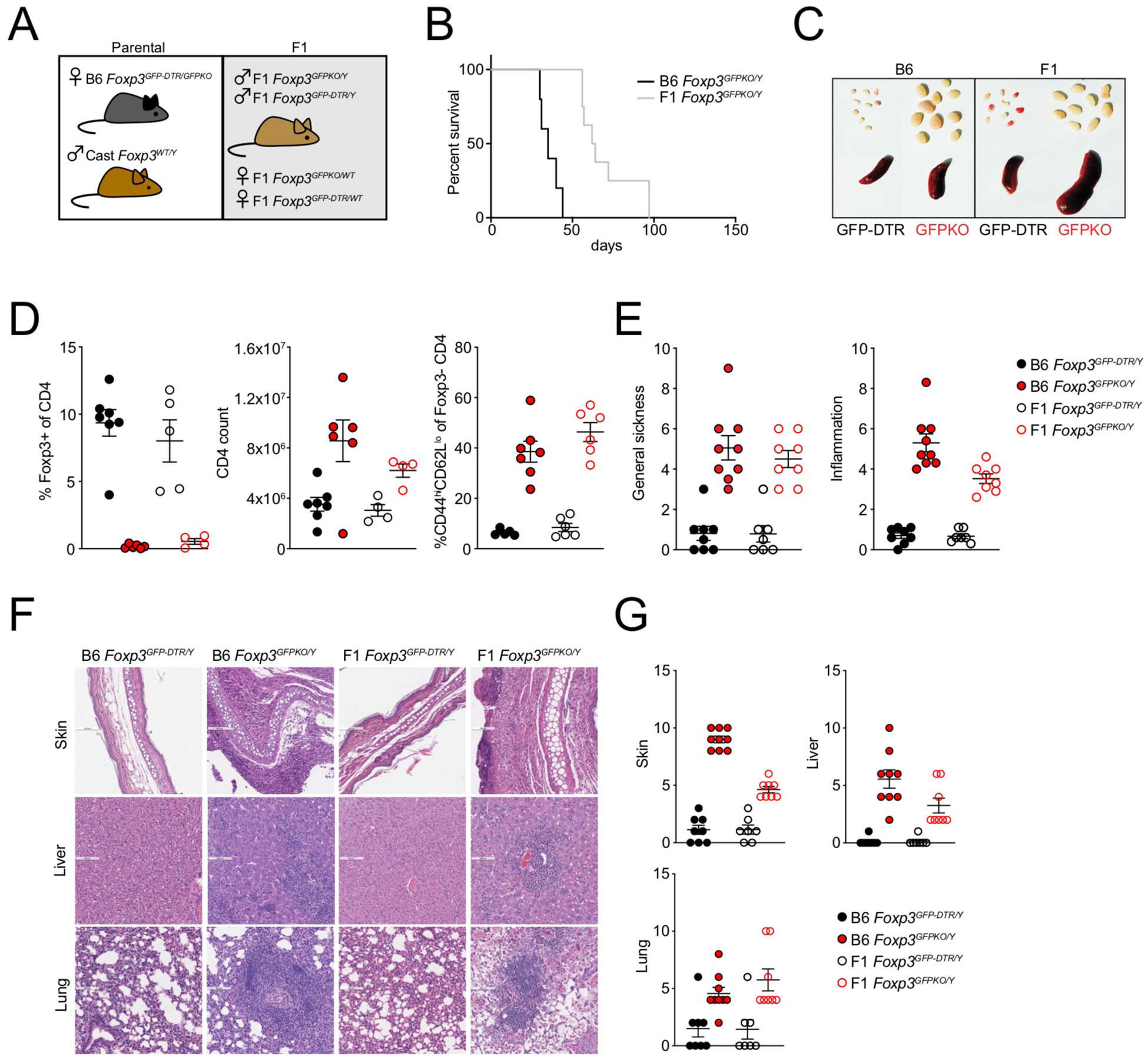
FIGURE 1: Foxp3-dependent suppression of autoimmune inflammation in B6/Cast F1 mice.

(A): Generation of experimental F1 mice. Female B6 Foxp3DTR-GFP/GFPKO mice were bred to male Cast/EiJ (Cast) mice.
(B): Survival of male Foxp3GFPKO/Y B6 (n=5) and B6/Cast F1 (n=8) mice.
(C): Spleens and lymph nodes of B6 and B6/Cast F1 Foxp3DTR-GFP and Foxp3GFPKO mice.
(D): CD4 T cell composition in lymph nodes of B6 and B6/Cast F1 Foxp3DTR-GFP and Foxp3GFPKO mice determined by flow cytometry. For Figures C-D, mice were analyzed at 3 weeks of age; data points were accumulated over multiple experiments.
(E): Sickness and inflammation scores based on combined histological assessment of skin, liver, and lung from B6 Foxp3DTR-GFP (n=8), B6/Cast F1 Foxp3DTR-GFP (n=7), B6 Foxp3GFPKO (n=9) and B6/Cast F1 Foxp3GFPKO (n=7) mice.
(F): H&E staining of skin, liver, and lung from B6 and B6/Cast F1 Foxp3DTR-GFP and Foxp3GFPKO mice.
(G): Pathology scores of individual tissues.
For E-G, data are derived from accumulated formaldehyde-preserved tissues from multiple experiments with 1–4 mice per group.
