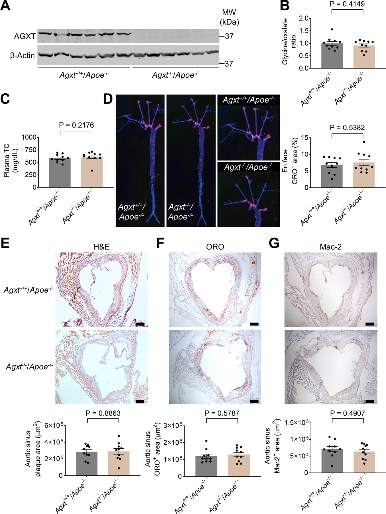
Figure 3. Oxalate homeostasis is maintained and atherosclerosis is unaltered in female Agxt−/−/Apoe−/− mice.

(A) Western blot analysis confirming the loss of AGXT in livers from female Agxt−/−/Apoe−/− mice (n=6).
(B) Glycine to oxalate ratio in plasma from female Agxt−/−/Apoe−/− and Agxt+/+/Apoe−/− mice (n=10).
(C-G) Female Agxt−/−/Apoe−/− and Agxt+/+/Apoe−/− mice were fed a WD for 12 weeks (n=10): (C) plasma total cholesterol (TC), (D) atherosclerosis in the aortic tree, (E) H&E staining, (F) Oil Red O staining, and (G) Mac2 immunohistochemistry of aortic sinus. (scale bar: 200 μm).
Unpaired t test for B, D, E and G. Mann-Whitney U test for C and F. Data are presented as mean ± SEM. All points and P values are shown.
