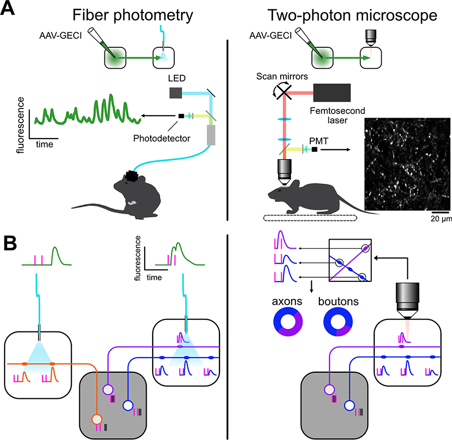
Fig. 1.
Differences between recordings of calcium signals in axons using fiber photometry and two-photon microscopy. A, Both methods allow recording activity in specific projections. Fiber photometry (left) in not image forming and is amenable to recordings during freely moving behaviors and from regions at any depth in the brain. Two-photon microscopy (right) results in an image of the recorded axons but it is limited to head-fixed animals and, unless tissue is removed, to superficial areas. B, Neurons in a given area can project to different targets. Their activity (black ticks) is locked to different behavioral events (magenta ticks). Calcium activity in axons in the target area reflects somatic activity and is correlated across boutons of the same axons, albeit with different amplitude. Left, fiber photometry-based recordings reflect the average population activity in the target region. Right, using two-photon microscopy, calcium activity can be extracted from individual boutons. Given the high correlation across boutons of the same axon, activity be quantified both in axons and boutons.

