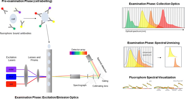
Figure 5.
Spectral Cytometry. Sample preparation is similar to that of conventional flow cytometry; detection antibodies conjugated with fluorophores are used for labeling. Within the flow cell, sample then enters the sample stream focused such that a single cell is interrogated by one or more lasers resulting in light dispersion and activation of the fluorophores. Emitted fluorescence is then collected by a detector array. Spectral overlap between multiple fluorophores on the same cell then separated using spectral unmixing, enabling the simultaneous detection of fluorophores with overlapping emission spectra (figure adapted from reference (12); emission spectra from www.cytekbio.com)

