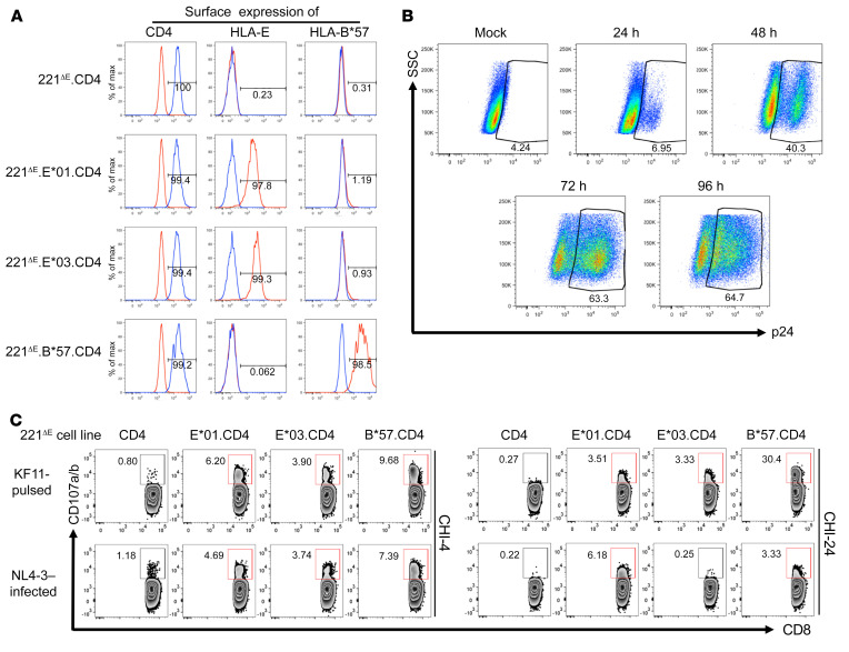
Figure 3. Activation of KF11-specific CD8+ T cells by HIV-1–infected target cells expressing HLA-E and CD4.
(A) Confirmation of surface expression of CD4 by 221ΔE.CD4, 221ΔE.E*01.CD4, 221ΔE.E*03.CD4, and 221ΔE.B*57.CD4 cell lines. Staining by isotype-matched negative control mAbs is also depicted in each case. (B) Percentages of p24+ cells and increasing intensity of cytoplasmic p24 staining (mAb Kc57) in a representative NL4-3–infected cell line (221ΔE.CD4) over 96 hours. (C) Degranulation of CD94–CD8hi T cells from patients CHI-4 and CHI-24 after incubation with NL4-3–infected 221ΔE.CD4, 221ΔE.E*01.CD4, 221ΔE.E*03.CD4, or 221ΔE.B*57.CD4 target cells. Effector cells were generated by in vitro stimulation with 10 μg/mL KF11 peptide. Target cells were assessed 48 hours after infection with 18% of the cells staining for p24. KF11-pulsed target cells were included as positive controls, while 221ΔE.CD4 cells (KF11-pulsed or NL4-3–infected) served as negative controls.

