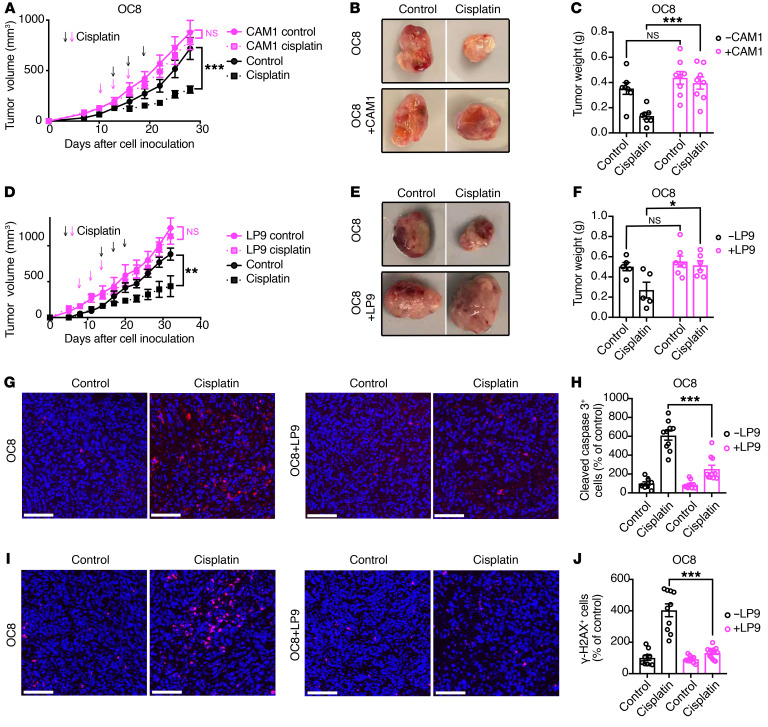
Figure 1. Cancer-associated mesothelial cells promote ovarian cancer platinum resistance.
(A–F) Effect of primary CAM1 (A–C) or LP9 (D–F) coinjection on cisplatin response of primary OC8 HGSOC cells in vivo. OC8 cells or OC8 cells plus LP9 or CAM1 mesothelial cells were injected subcutaneously into female immunodeficient mice and treated with or without cisplatin every 3 days for 3 cycles. Tumor growth curves are shown in A (n = 7–8 mice per group) and D (n = 5–7 mice per group). Representative xenograft images are shown in B and E. Xenograft weights at the end point are shown in C and F. Arrows show scheme of cisplatin treatment: magenta arrows for mesothelial cell–coinjected groups, black arrows for OC8 cell alone groups. (G–J) Representative images and quantification of cleaved caspase-3 (G and H) and γ-H2AX (I and J) immunofluorescence staining in OC8 and LP9 coinjected tumors. Scale bars: 100 μm. Quantification of positive cells (percentage of control) is based on 10 random fields from more than 3 tumors in each group. Each dot represents 1 field. Nuclei were stained with DAPI (blue). Data are presented as mean ± SEM. *P < 0.05; **P < 0.01; ***P < 0.001, 2-way ANOVA (A, C, D, and F) and 2-tailed Student’s t test (H and J).

