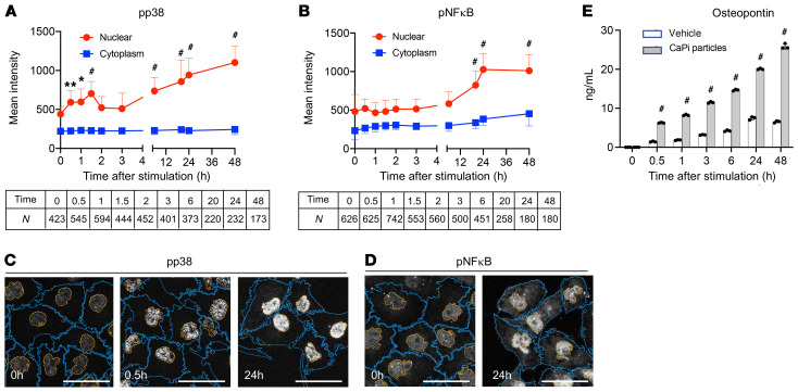
Figure 4. Calcium phosphate particles activate the p38/NF-κB pathway and induce osteopontin secretion.
(A and B) HK-2 cells were incubated with synthesized calcium phosphate particles (10 μg phosphorus/mL) for the indicated durations and subjected to immunocytochemical analysis using antibodies against p-p38 (A) or p–NF-κB (B). The confocal microscopic images were analyzed using Nikon NIS-Elements software to determine the intensity of the fluorescence signals from the nucleus (red) and cytoplasm (blue). Data are indicated as the mean ± SD. N = the number of cells analyzed for each time point. *P > 0.25 (effect size) and **P > 0.3 (effect size); #P > 0.35 (effect size) versus time 0 (without stimulation with calcium phosphate particles), by Brunner-Munzel test. (C and D) Representative confocal images of immunocytochemistry for p-p38 (C) and p-NF-κB (D) are shown. Plasma membranes are outlined in orange and nuclear membranes in blue. Scale bars: 50 μm. (E) The osteopontin concentration was measured by ELISA in conditioned medium of HK-2 cells incubated with or without calcium phosphate particles (10 μg phosphorus/mL) for the indicated durations. n = 3 for each column. #P < 0.0001 versus vehicle, by 2-way ANOVA with Šidák’s multiple-comparison test.

