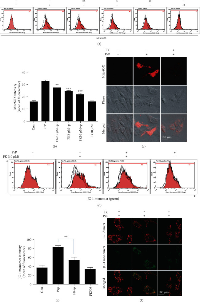
Figure 7.

PrP (106-126)-mediated calcineurin activation induced neurotoxicity via mitochondrial dysfunction. SK-N-SH cells were pretreated with FK506 (1 h) and then exposed to 100 μM PrP (106-126) for 6 hours. (a) Mitochondrial ROS was evaluated by a MitoSOX assay. (b) Bar graph showing the averages of the red fluorescence (MitoSOX). Values represent the mean ± SEM (n = 10). ∗∗p < 0.01, ∗∗∗p < 0.001 vs. PrP. (c) MitoSOX fluorescence images were obtained after exposure to 100 μM PrP (106-126) (6 h) in the absence or presence of FK506 (10 μM, 1 h). (d) Mitochondrial membrane potential was evaluated by a JC-1 assay using flow cytometry. In green fluorescent colors, JC-1 accumulates as green monomers in the mitochondria of cells with impaired mitochondrial membrane potential function. (e) Bar graph showing the averages of the green fluorescence (JC-1 monomers). Values represent the mean ± SEM (n = 10). ∗∗p < 0.01 vs. PrP. (f) JC-1 fluorescence images were obtained after exposure to 100 μM PrP (106-126) (6 h) in the absence or presence of FK506 (10 μM, 1 h).
