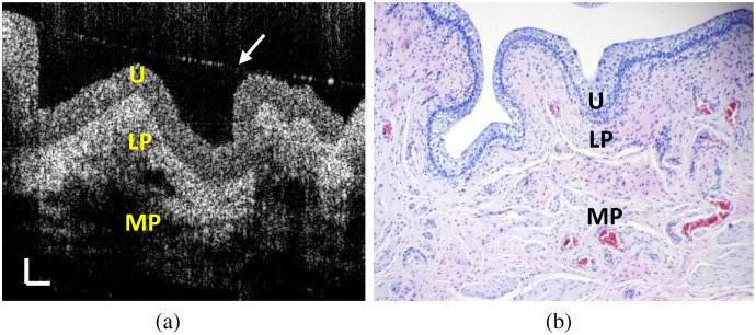
Fig. 1.
OCT images of a fresh porcine bladder tissue sample under a stretched condition. (a) A normal OCT image, (b) corresponding H&E histological image (), and (c) 100 OCT images captured at . The arrow in (a) marked the interface of a thin water layer over the tissue surface. U, urothelium; LP, lamina propria (LP); MP, muscularis propria (scale bar: 0.1 mm) (Video 1, MP4, 2 MB [URL: https://doi.org/10.1117/1.JBO.26.8.086002.1]).

