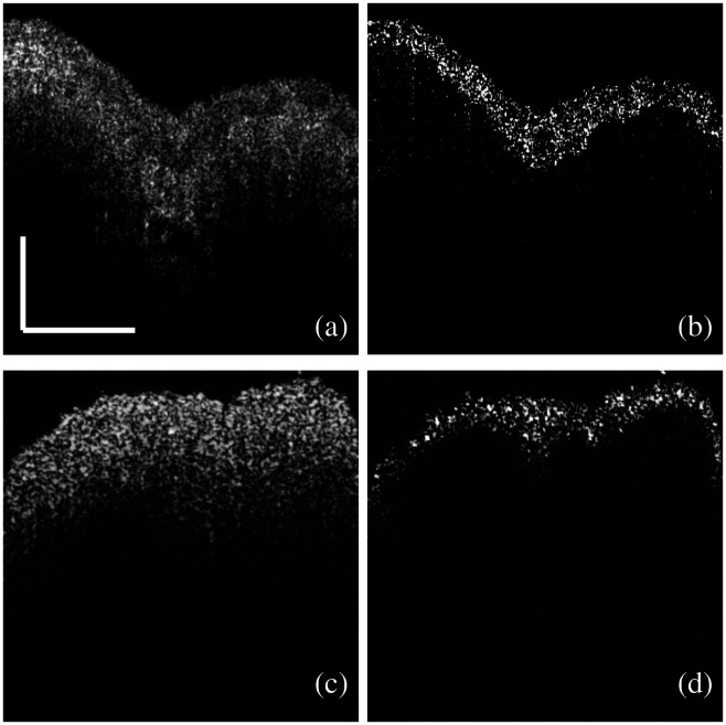
Fig. 5.
Segmentation of the urothelium with IM. (a) An OCT image of a fresh porcine bladder tissue sample with significant stretching. (b) The image reconstructed after the subtraction of the OCT complex signals between the two sequential images like (a). (c) An OCT image of a fresh porcine bladder tissue sample with minimal stretching. (d) The image reconstructed after the subtraction of the OCT complex signals between the two sequential images like (c) (scale bar: 0.5 mm).

