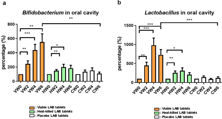
Fig. 1.
a Measurement of Bifidobacterium populations on the oral mucosal surfaces. V viable probiotic tablets; H heat-killed inactivated probiotic tablets; C placebo tablets (control); and LAB lactic acid bacteria. Collection and analysis of salivary samples at Weeks 0, 2, 4, and 6 were performed. Data were presented as the mean ± standard deviation (SD). Two-tailed t-test was used to analyzed statistical difference. Statistical significances were marked with *P < 0.05, **P < 0.01, ***P < 0.001. b Measurement of Lactobacillus populations on the oral mucosal surfaces. V viable probiotic tablets; H heat-killed inactivated probiotic tablets; C placebo tablets (control); and LAB lactic acid bacteria. Collection and analysis of salivary samples at Weeks 0, 2, 4, and 6 were performed. Data were presented as the mean ± standard deviation (SD). Two-tailed t-test was used to analyzed statistical difference. Statistical significances were marked with *P < 0.05, **P < 0.01, ***P < 0.001

