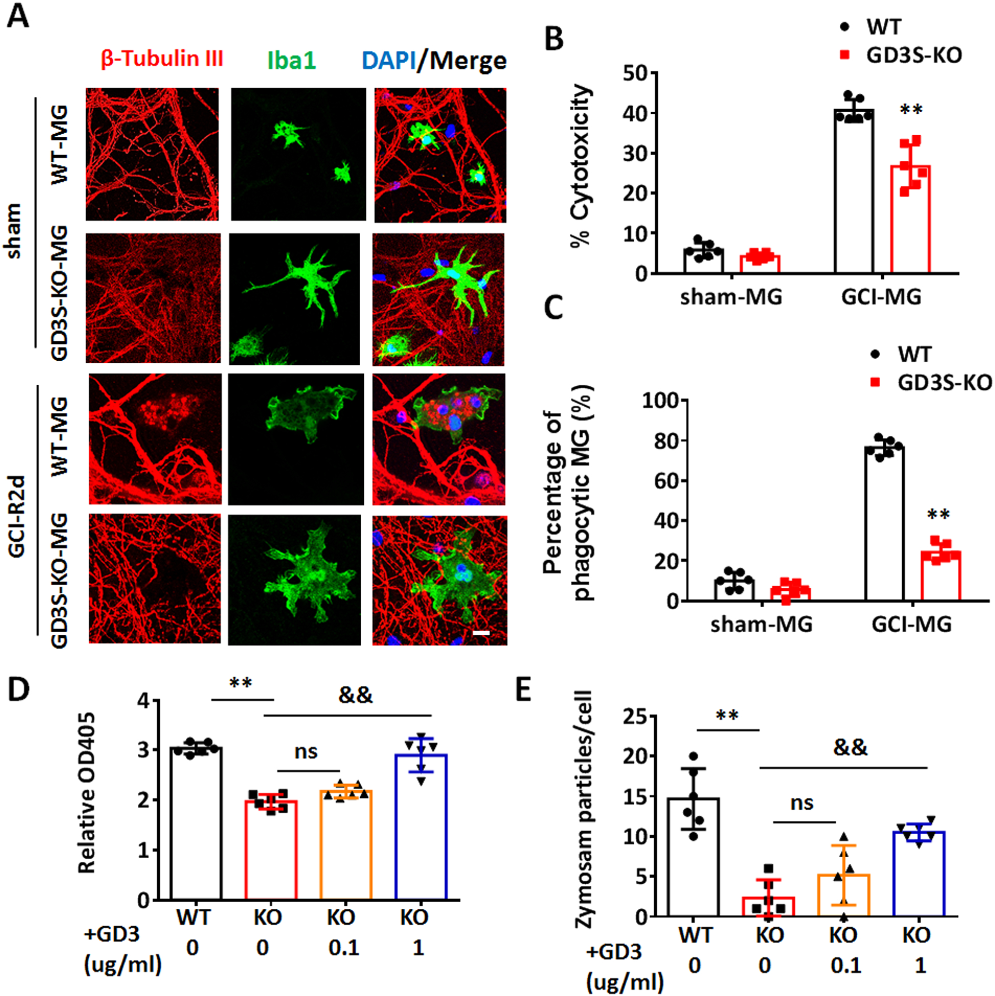
Figure 8. Evidence that ganglioside GD3 regulates the neurotoxicity and phagocytic capacity of microglia in vitro.

A. Representative images of primary hippocampal neuron (β-tubulin III, red) co-cultured with microglia (MG) (Iba1, green) from sham or GCI-R2d mouse brain for 72 h. B. Neurotoxicity of co-cultured microglia was measured by LDH assay compared between the control wells without microglia co-culture. C. The percentage of phagocytic MG was quantified by counting the number of MG with engulfed neuronal elements and dividing that by the total number of MG in 10 randomized fields in each well. D. Phagocytic capacity of primary microglia isolated from GCI-R2d mouse brain, with/without exogenous GD3 treatment were measured by Zymosan substrate assay at 48 hour in vitro. E. Phagocytic capacity of primary microglia isolated from GCI-R2d mouse brain, with/without exogenous GD3 treatment were measured by quantification of the average Zymosan particles number in each cell. Cells from 2 randomly captured images of each well were quantified. For the each experiments, n=6 mice/group. 2 mouse brains were pooled for isolating 1 sample of microglia for in-vitro culture. 2 independent cell cultures were performed for the neurotoxicity and phagocytic capacity assay, and data from triplicated wells were collected. **p<0.01 GD3S-KO vs. WT. &&p<0.01 vs. KO (0 ug/ml GD3). ns, not significant. Two-way ANOVA followed by post-hoc. Scale bar: 5 μm.
