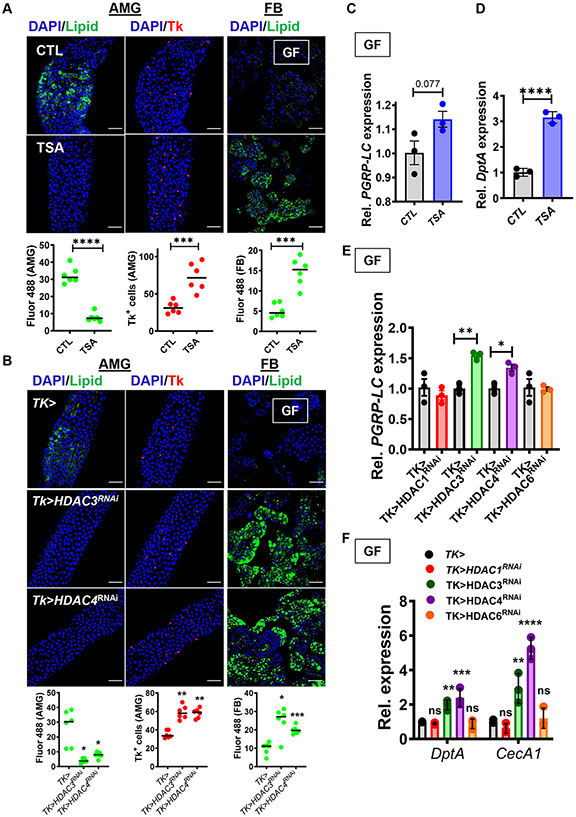
Fig 4: Inhibition of protein deacetylation inhibits accumulation of TAG and activates IMD pathway signaling in the GF fly intestine.
Representative micrographs (above) and quantification (below) of Bodipy staining (Lipid) and Tk immunofluorescence in the AMG and FB of (A) GF yw flies fed LB broth alone (CTL) or supplemented with trichostatin A (TSA), and (B) GF Tk> and Tk>HDAC3RNAi and Tk>HDAC4RNAi flies. This data was acquired in tandem with the data presented in Fig S5C and uses the same driver control. RT-qPCR quantification of (C) PGRP-LC and (D) DptA in GF yw flies fed LB broth alone (CTL) or supplemented with trichostatin A (TSA). RT-qPCR quantification of (E) PGRP-LC and (F) DptA and CecA1 in GF Tk> flies or with Tk> specific HDAC RNAi as noted. The mean measurement is indicated. Error bars represent the standard deviation. A student’s t-test (A, C, D), a Brown-Forsythe ANOVA with a Dunnett’s T3 multiple comparisons test (B) or an ordinary one-way ANOVA with Dunnett’s multiple comparisons test (E) was used to evaluate significance. ns not significant, * p<0.05, ** p<0.01, *** p<0.001, **** p<0.0001. See also Fig S5.

