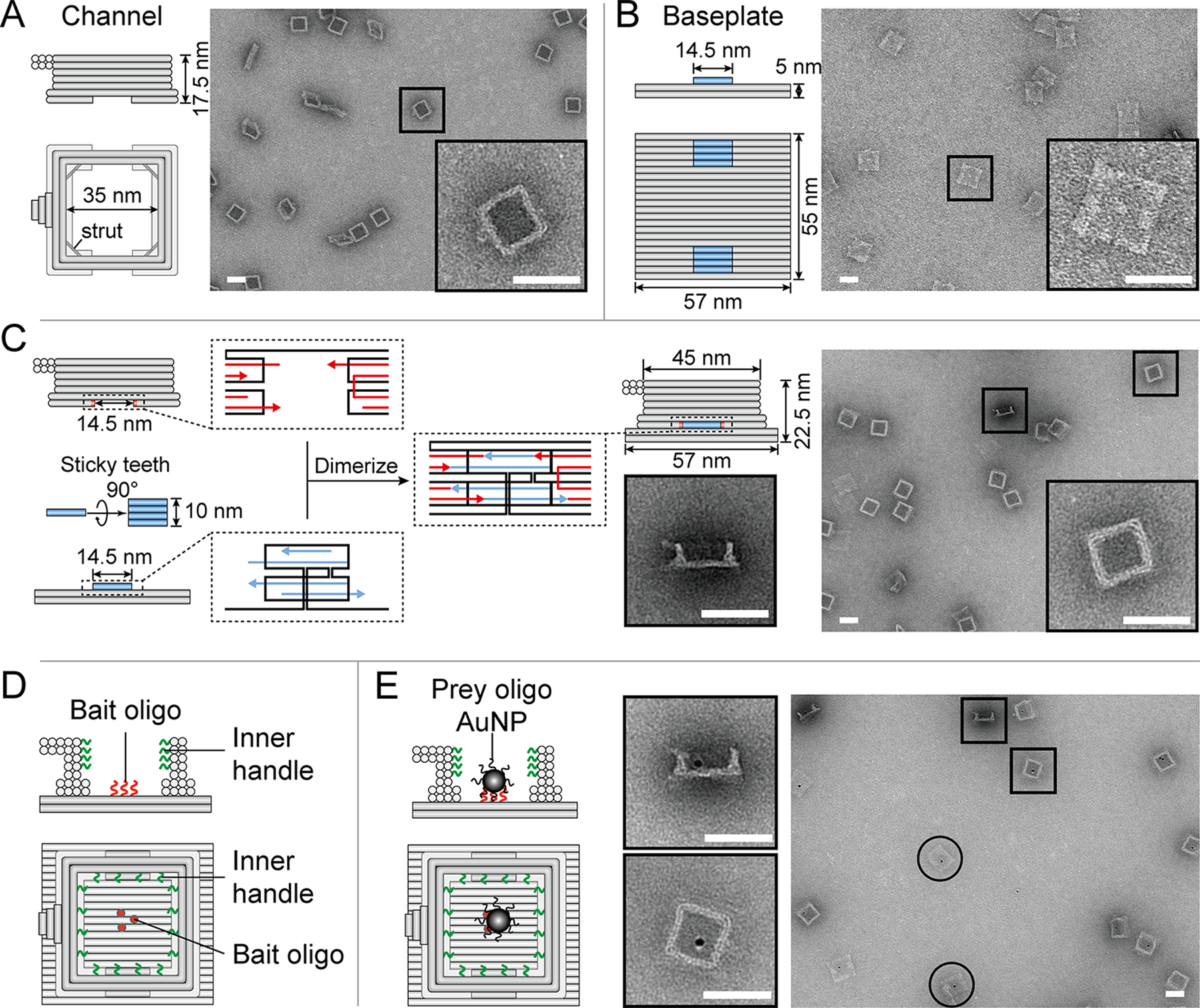
Figure 1.

A DNA-origami NanoTrap built from two pre-assembled parts, a channel and a baseplate. (A) Cartoon models and negative-stain EM images of the channel. (B) Cartoon models and negative-stain EM images of the baseplate. (C) Two teeth (blue) with sticky ends (blue arrows) mediate the baseplate attachment onto the channel, which contains two cavities with complementary sticky ends (red arrows), resulting in the formation of a NanoTrap (cartoon model and negative-stain EM images shown on the right). (D) Up to four layers of DNA handles (12 handles per layer) protrude from the channel wall (green curls), serving as anchor points for anti-handle-conjugated proteins. The baseplate displays three “bait” oligonucleotides (red curls/dots) to capture nanoscale objects carrying “prey” oligonucleotides. (E) Cartoon models and EM images showing the immobilization of prey-oligo-modified AuNPs (dark spots) inside the NanoTrap. Circles indicate standalone baseplates. Scale bars = 50 nm.
