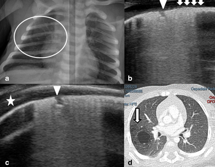
Fig. 2.
a Chest X-ray showing a suspected hyperlucent ovalar lesion in the right hemy-thorax (white circle); b, c post-natal lung ultrasound shows a characteristic thickened pleura with microcistic-like hypoechoic lesions within the pleural line (white triangles), a bigger round subpleural lesion (white arrow) and posterior vertical artifacts. The close parts of the pleural line is normal (white star); d the CT scan confirmed the lung malformation (arrow)

