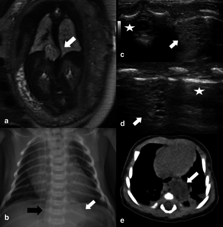
Fig. 3.
a Prenatal MRI showing a thoracic mass with deviation of the aorta (white arrow). b Chest X-ray showed a suspected left supra diaphragmatic lesion (white arrow) and deviation of the aorta through the right (black arrow); c post natal lung ultrasound (intercostal/transvers view) shows an echogenic lesion, having inside multiple hypoechoic microcystic lesions (white arrow), close to the vertebrae (white star); d lung ultrasound (longitudinal view) shows the same lesion (white arrow) surrounded by normal pleural line (white star); e CT scan confirmed the presence of the thoracic lesion (white arrow)

