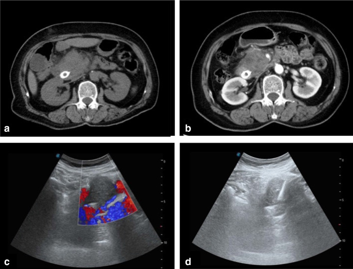
Fig. 5.
CT and US of a 65-year-old man with a pancreatic head mass detected after biliary stent implantation. The pathology diagnosis of the mass was pancreatic ductal adenocarcinoma. a Transverse CT image showing iso-density of the pancreatic head mass. b Contrast-enhanced CT showing a low-intensity tumor in the pancreatic head. c, d US-guided puncture of the pancreatic head hypoechoic mass

