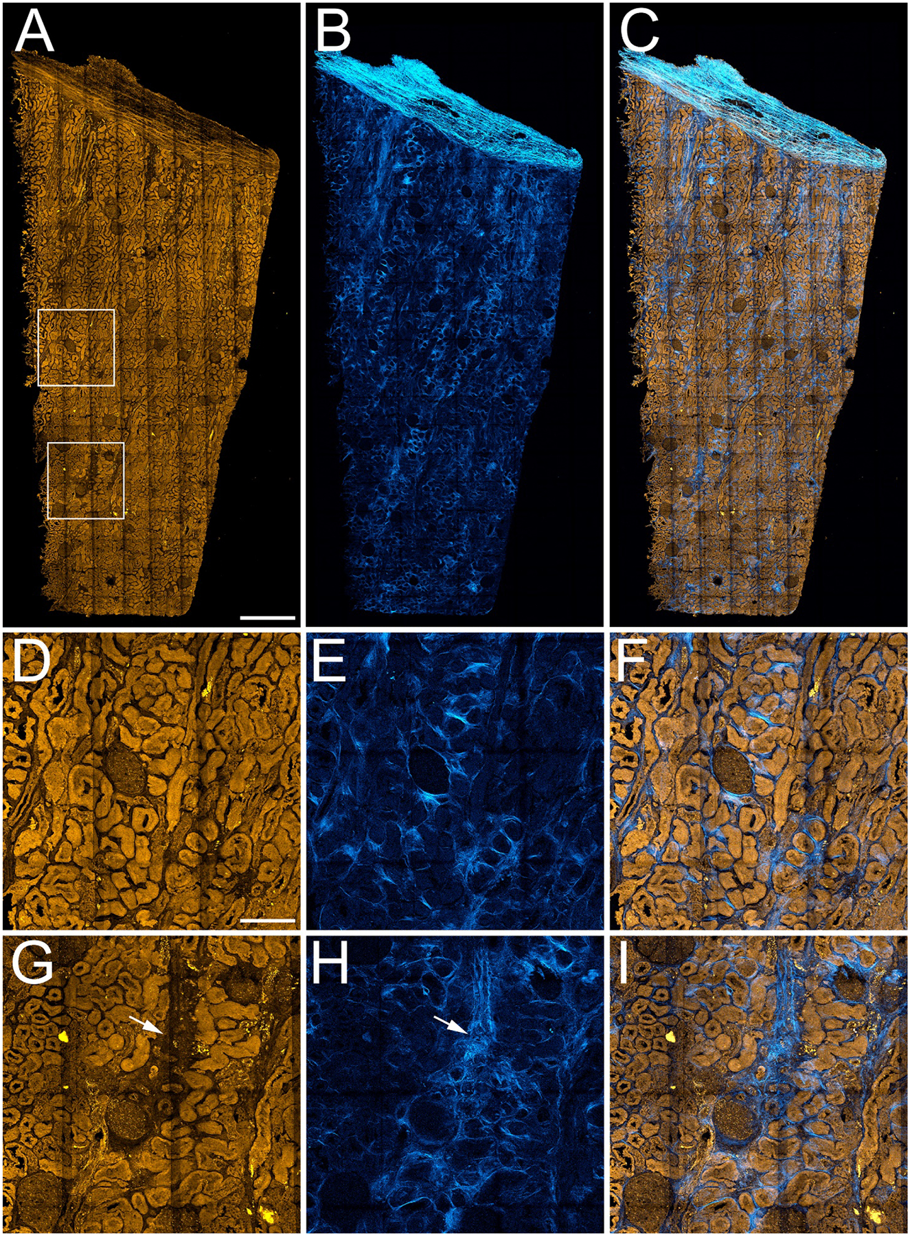
Figure 2. 3D multiphoton microscopy of unlabeled nephrectomy.

Mosaic of high-resolution image volumes collected from a 4 mm by 9 mm, 50 micron thick section of paraformaldehyde-fixed human nephrectomy tissue. Panel A – Maximum projection image of 3D volume of tissue autofluorescence. Panel B- Maximum projection of 3D volume of second harmonic generation (SHG) images. Panel C – Overlay of autofluorescence and SHG. Panels D-F show corresponding 4X magnification images of the region indicated in the upper box in panel A, and panels G-I show corresponding 4X magnification images of the region indicated in the lower box in panel A. Arrows in panels G and H indicate regions of apparent tubular dropout and fibrosis. Scale bar in panel A represents 1 mm. Scale bar in panel D represents 250 microns in D-I.
