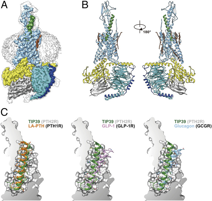
Fig. 1.
The overall cryo-EM structure of the TIP39–PTH2R–Gs complex. (A) Cut-through view of the cryo-EM density map that illustrates the TIP39–PTH2R–Gs complex and the disk-shaped micelle. The unsharpened cryo-EM density map at the 0.06 threshold shown as gray surface indicates a micelle diameter of 11 nm. The colored cryo-EM density map is shown at 0.12 threshold. (B) Model of the complex as a cartoon, with TIP39 as helix in green. The receptor is shown in blue, Gαs in yellow, Gβ subunit in cyan, Gγ subunit in navy blue, and Nb35 in gray. (C) The binding pocket of PTH2R accommodates peptide ligands of class B1 receptors. TIP39 is compared with LA-PTH (Left), GLP-1 (Middle), and glucagon (Right), respectively

