Fig. 1.
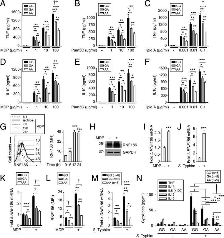
Human MDMs from rs6426833 A risk carriers demonstrate reduced PRR-induced cytokines and RNF186 expression. (A–F) MDMs from rs6426833 GG, GA, or AA carriers (n = 10 donors/genotype) were treated for 24 h with 1, 10, or 100 µg/mL MDP (recognized by NOD2) (A and D); 1, 10, or 100 µg/mL Pam3Cys (recognized by TLR2) (B and E); or 0.001, 0.01, or 0.1 µg/mL lipid A (recognized by TLR4) (C and F). TNF (A–C) and IL10 secretion (D–F) stratified by rs6426833 genotype. (G–I) MDMs were treated with 100 μg/mL MDP. (G) RNF186 protein expression at the indicated times. (Left) Representative histogram with mean fluorescence intensity (MFI) values. Isotype control is from 24-h treated cells. (Right) Summary graph (n = 4 donors; similar results in additional n = 4). (H) RNF186 expression at 24 h assessed by Western blot. (I) RNF186 mRNA expression at 4 h (n = 4 donors; similar results for an additional n = 4). (J) MDMs (n = 5 donors; similar results in additional n = 4) were cocultured with S. Typhimurium. RNF186 mRNA at 4 h. (K and L) MDMs (n = 10 donors/genotype; similar results in additional n = 6/genotype) were treated with 100 μg/mL MDP. (K) RNF186 mRNA at 4 h. (L) RNF186 protein expression at 24 h. (M and N) Human intestinal myeloid cells were cocultured with S. Typhimurium. RNF186 mRNA at 2 h (M) and cytokine secretion at 12 h (N) stratified on rs6426833 genotype. Mean + SEM. NT, no treatment. *P < 0.05; **P < 0.01; ***P < 0.001; †P < 1 × 10⁻⁴; ††P < 1 × 10⁻⁵.

