Fig. 4.
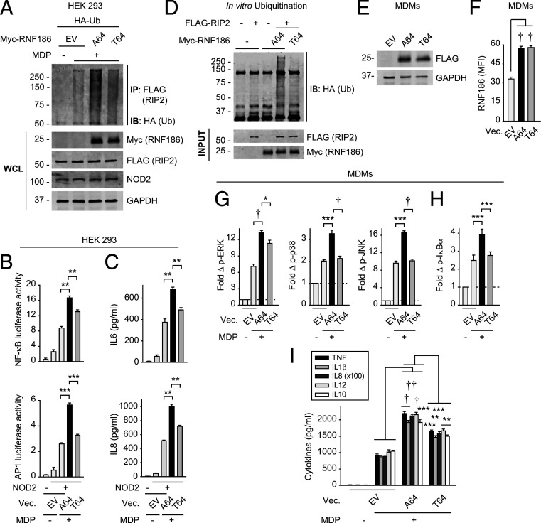
The rare RNF186 A64T IBD-risk variant results in decreased NOD2-induced outcomes compared to WT RNF186. (A) HEK293 cells were transfected with EV, myc-RNF186-A64 (WT) or myc-RNF186-T64 (rare risk variant), and FLAG-RIP2, NOD2, and HA-ubiquitin. Cells were then treated with 100 μg/mL MDP for 45 min. RIP2 (FLAG) was IP and the association of ubiquitinated proteins was assessed by α-HA (IB). WCL were assessed for equal loading. Representative of three independent experiments. (B and C) HEK293 cells were transfected with EV, RNF186-A64 (WT), or RNF186-T64 (risk variant) along with NOD2 ± AP-1 or NFκB luciferase and Renilla constructs. Cells were then treated with 100 μg/mL MDP (representative of two independent experiments). NFκB and AP-1 luciferase activity at 6 h (four replicates) (B) and cytokine secretion at 24 h (five replicates) (C). (D) In vitro ubiquitination was assessed with purified HA-ubiquitin ± purified FLAG-RIP2 ± purified myc-RNF186-A64 or myc-RNF186-T64. Ubiquitin protein (anti-HA) was detected by Western blot. Representative of three independent experiments. (E–I) MDMs from rs6426833 AA risk carriers (low RNF186-expressors) were transfected with EV, FLAG-tagged RNF186-A64 (WT), or RNF186-T64 (risk variant). (E and F) RNF186 protein expression by Western blot (E) or flow cytometry (F) (n = 6 donors). (G–I) Cells were treated with 100 μg/mL MDP (n = 6). (G and H) Fold phospho-proteins at 15 min. (I) Cytokines at 24 h. Mean + SEM. Vec, vector. *P < 0.05; **P < 0.01; ***P < 0.001; †P < 1 × 10⁻⁴; ††P < 1 × 10⁻⁵.

