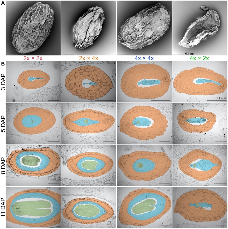
Figure 2.
Seed development of parental and reciprocal hybrid seeds. A, Scanning electron micrograph images of a representative seed from each cross (M. guttatus—2x × 2x [red]; 2x × 4x [orange]; M. luteus—4x × 4x [blue]; 4x × 2x [green]) is displayed. B, Histological sections were made of each cross through a developmental progression from 3, 5, 8, to 11 DAP. Crosses are displayed in columns and DAP is displayed in rows. Within the images, the seed coat is orange, embryo is green, and endosperm is blue. Scale bars, 0.1 mm

