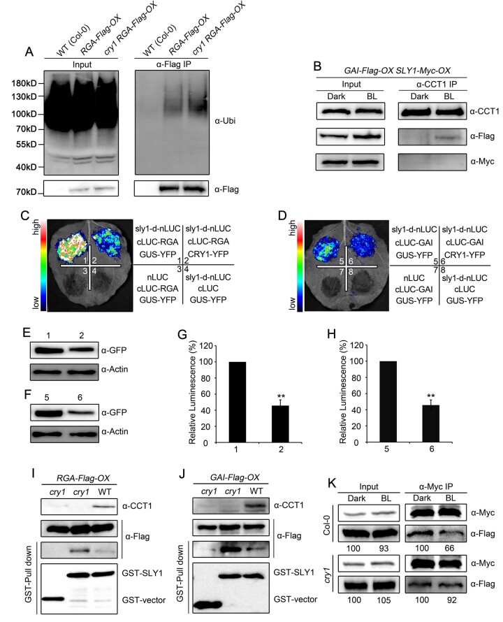
Figure 4.
CRY1 impairs the interaction of DELLA proteins with SLY1. A, In vivo ubiquitination assays showing CRY1-mediated inhibition of RGA ubiquitination in blue light. Seedlings for WT(Col-0), RGA-Flag-OX in WT and cry1 were grown in blue light (50 μmol·m–2·s–1) for 5 days, and then treated with 100-μM GA3 and 50-μM MG132 in liquid MS medium for 2 h, followed by immunoprecipitation with anti-Flag beads. The IP signals (RGA-Flag) were detected in immunoblots probed with anti-ubiquitin, and input signals were detected with anti-Flag antibodies. Two independent experiments were performed, and one is shown. B, Co-IP assays showing no interaction of CRY1 with SLY1 in Arabidopsis. Dark-adapted double transgenic GAI-Flag-OX/SLY1-Myc-OX seedlings were kept in darkness or exposed to blue light (BL, 50 μmol·m–2·s–1) for 1 h, followed by immunoprecipitation with anti-Myc beads. The IP (CRY1) and co-IP signals (SLY1 and GAI) were detected in immunoblots probed with anti-Myc and anti-Flag antibodies, respectively. Two independent experiments were performed, and one is shown. C–H, Split-luciferase complementation imaging assays indicating that CRY1 prevents the interaction between SLY1 with RGA (C) and GAI (D) in N. benthamiana leaves. The quantification of luciferase activity for the samples in (C) and (D) is shown in (G) and (H), respectively. Data are shown as means of biological triplicates ± sd (n = 4; Student’s t test, **P < 0.01). The accumulation of CRY1-YFP and GUS-YFP (negative control) was detected with an anti-GFP antibody in (E) and (F). I, J, Cell-free GST pull-down assays showing that CRY1 prevents the interaction between SLY1 and RGA (I) or GAI (J). GST–SLY1 served as bait. Preys were protein extracts prepared from dark-adapted RGA-Flag-OX GAI-Flag-OX (WT and cry1 backgrounds) seedlings exposed to blue light (50 μmol·m–2·s–1) for 1 h. Two independent experiments were performed, and one is shown. K, Co-IP assays showing that CRY1 prevents the interaction of SLY1 with GAI in Arabidopsis. Total protein was extracted from dark-adapted double transgenic seedlings co-expressing SLY1-Myc and GAI-Flag in the WT and cry1 backgrounds and exposed to blue light (50 μmol·m–2·s–1) for 1 h, followed by immunoprecipitation with anti-Myc beads. The immunoprecipitates were probed with anti-Flag and anti-Myc antibodies. Relative band intensities were normalized for each panel and are shown below each lane. Two independent experiments were performed, and one is shown.

