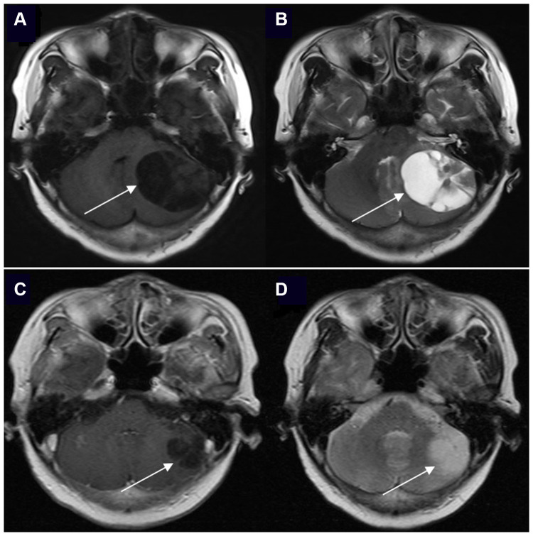
Figure 1.
The brain enhanced MRI of a 61-year-old woman with non-small cell lung cancer and EGFR 21L858R mutation before and after EGFR-TKI treatment. The positions indicated by the arrows are the lesions. (A) Contrast-enhanced T1-weighted MR image at baseline. (B) Contrast-enhanced T2-weighted MR image at baseline. (C) Contrast-enhanced T1-weighted MR image after icotinib (first-generation EGFR-TKI) treatment and whole brain radiotherapy (prescription dose and fraction schedule of 30Gy/10F) treatment for 1 month. (D) Contrast-enhanced T2-weighted MR image after icotinib and whole brain radiotherapy treatment for 1 month.

