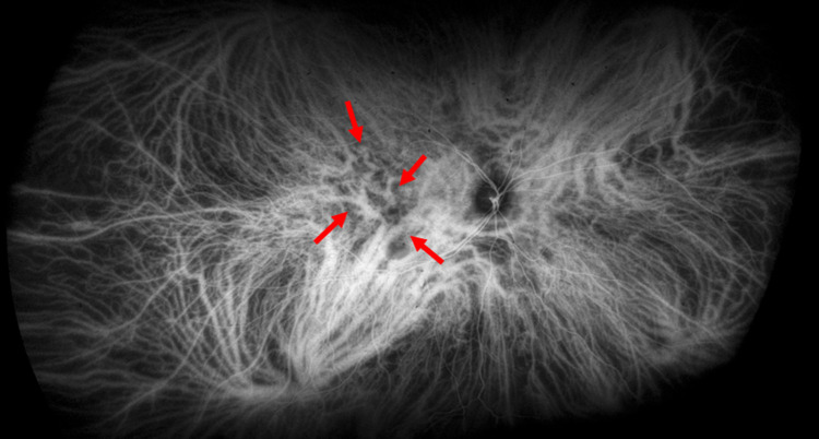
Figure 4.
Early-mid phase ultra-widefield indocyanine green angiography image of the right eye of a 38-year-old man with complex central serous chorioretinopathy. There is marked congestion of choroidal veins that appear abnormally enlarged, especially in the inferotemporal and superonasal vortex vein systems. Note the dense network of inter-vortex venous anastomoses in the macular and peripapillary region connecting the superonasal, superotemporal and inferotemporal vortex vein systems (red arrows).

