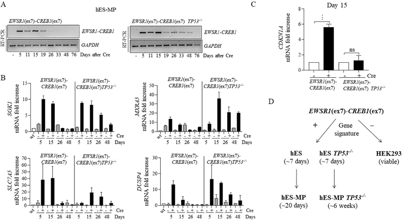
Figure 6. Prolonged viability of hES-derived mesenchymal cells expressing EWSR1-CREB1 in TP53−/− background.
(A) Time course in days for EWSR1-CREB1 fusion expression and (B) AFH core gene signature in wild type and TP53−/− genetic background. Histogram represents the fold increase to wild type (-Cre condition) of 1 representative experiment (n=2).
(C)CDKN1A expression is measured 15 days after the induction of the EWSR1-CREB1 fusion in wild type and TP53−/− cells. Histogram represents the fold increase of the +Cre condition compared to the corresponding -Cre condition and the error bars the standard deviation from the mean of 3 independent experiments. Statistical significance is calculated with a paired t-test comparing the +Cre with the corresponding -Cre condition. ***p<0.001, ns=not significant. (D) EWSR1(ex7)-CREB1(ex7) fusion expression impairs the cell proliferation/viability of hES cell after 7 days, regardless of TP53 deletion. On the contrary, in hES-MP the viability is prolonged and is further expanded by deletion of TP53 (see discussion for details).

