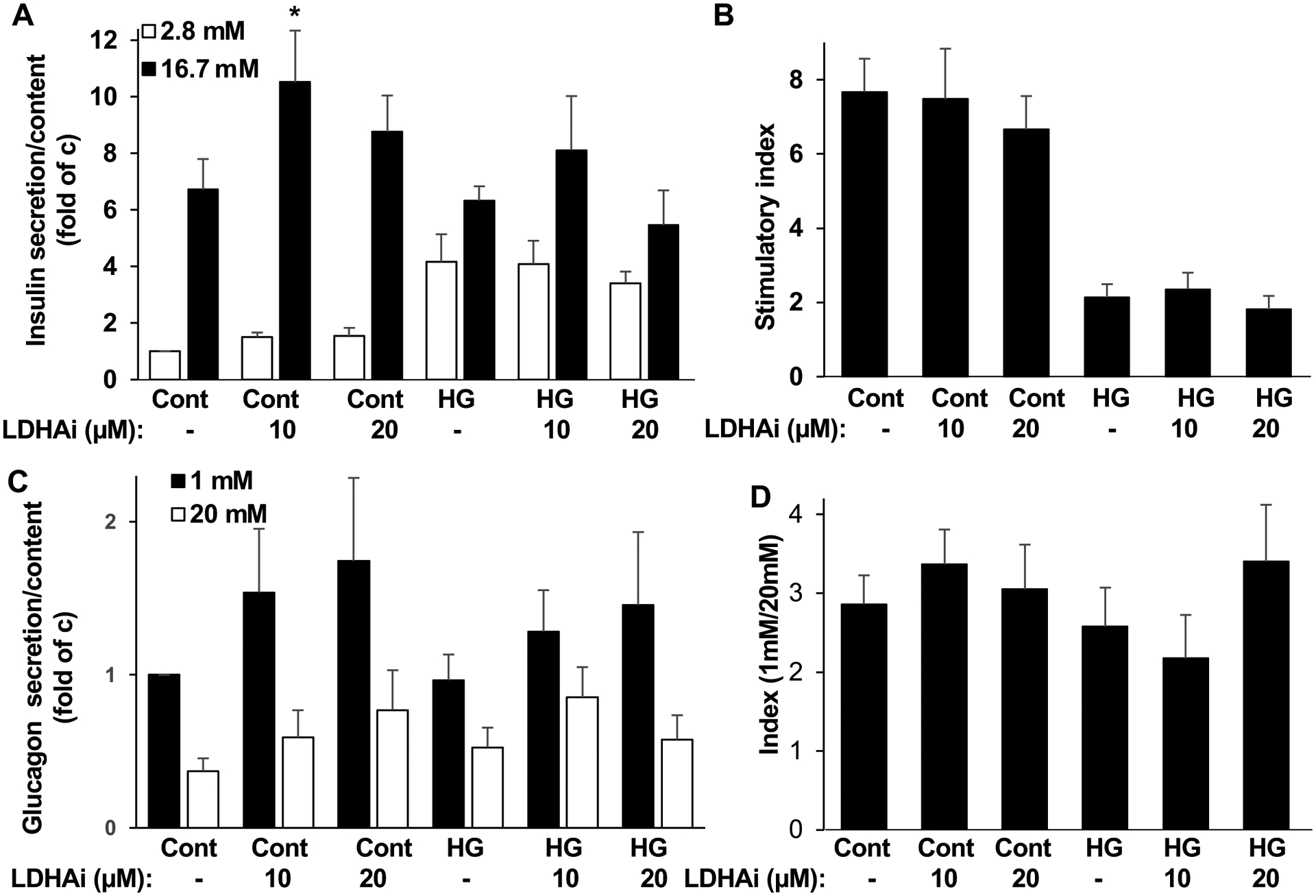
Figure 3. Impact of LDHA inhibition on insulin and glucagon secretion.

Isolated human islets treated with or without (control: cont) 10 or 20 μM LDHA inhibitor GSK2837808A (LDHAi) 1 hour before and during the 48 hour exposure to physiological glucose (5.5 mM; cont) or to high glucose (22 mM; HG). Thereafter, (A) Insulin secretion was analyzed during 1-h incubation with 2.8 mM (basal) and 16.7 mM (stimulated) glucose normalized to insulin content, (B) the insulin stimulatory index denotes the ratio of secreted insulin during 1-h incubation with 16.7 mM to secreted insulin at 2.8 mM glucose. (C) In a parallel set of islets, glucagon secretion was analysed during 1-h incubation with 1 mM and 20 mM glucose normalized to glucagon content, (D) the glucagon secretory index denotes the ratio of secreted glucagon during 1-h incubation with 1 mM to secreted glucagon at 20 mM glucose. A-D (n=8–9; from 3 different human islet isolations). Data are expressed as means ± SEM. *p<0.05 compared to untreated stimulated control.
