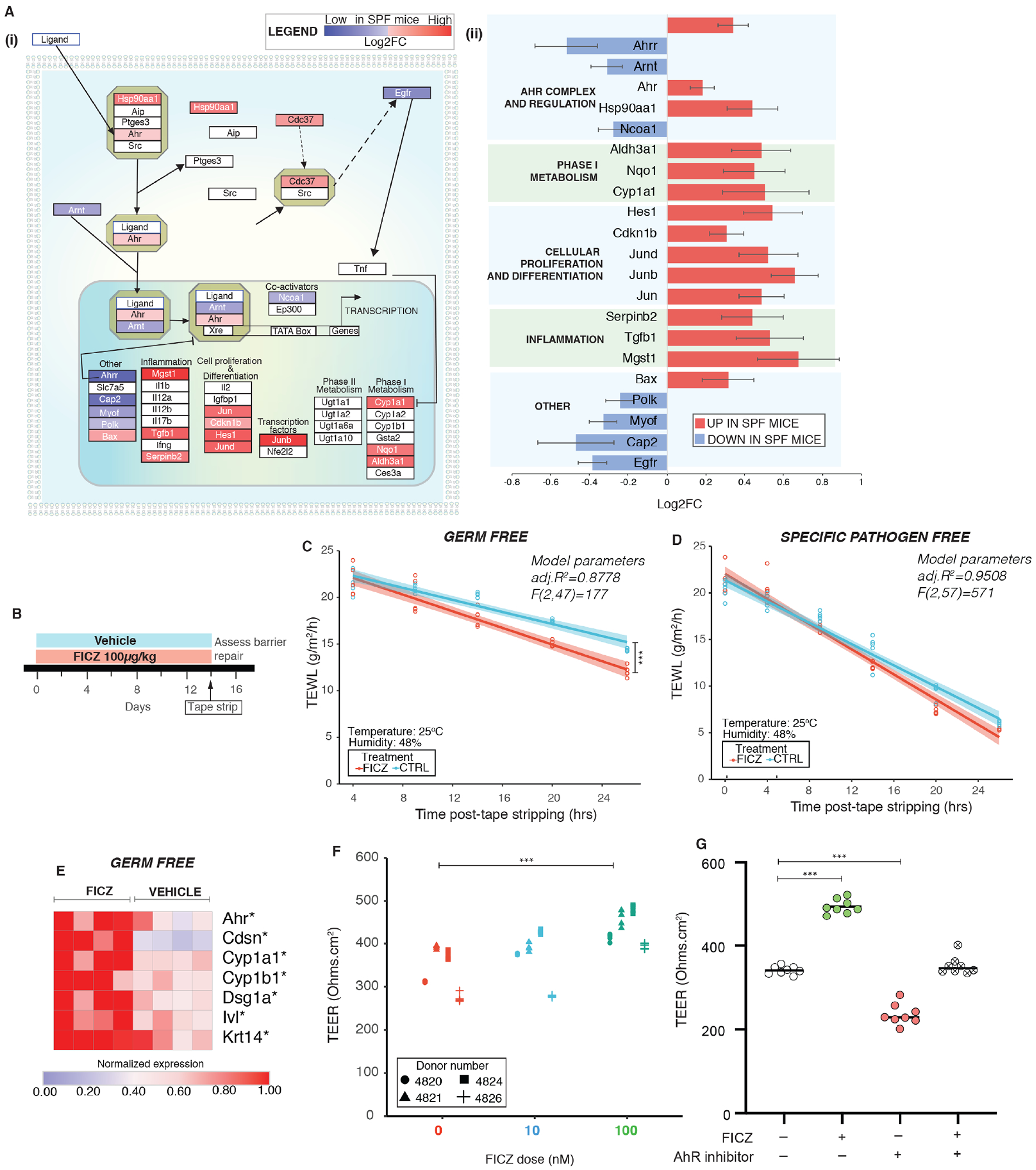
Figure 3. Activation of AHR signaling in skin rescues barrier dysfunction in germ free mice.

(A) Differentially expressed genes (DEGs) in SPF vs GF mice from RNAseq were (i) mapped onto the AHR pathway and (ii) Log2FC of significant DEGs (P<0.01) in SPF mice are represented. (B) Schematic illustrating experimental design. TEWL vs time readings were fitted by linear modeling and covariance was assessed by ANCOVA. Barrier recovery was compared in (C) GF mice [F (1,47) =21.9, ***P<0.001)] and (D) SPF mice [F(1,57)=2.98, *P=0.0492] that were either treated with FICZ or vehicle. (E) Expression of genes was assessed by qRT-PCR in GF skin treated with FICZ or vehicle (4 mice per group). *P<0.05, by T-test adjusted by Bonferroni correction. (F) Primary human keratinocytes grown on transwells in presence of FICZ (0nM, 10nM and 100nM) for three days and TEER was measured. Cells from different donors are represented by different symbol. See Figure S4. (G) Primary human keratinocytes were treated as indicated with FICZ and/or AHR inhibitor at 100nM doses. TEER values at the end of three days of treatment are reported. ***P<0.001 by T-test for panels F and G.
