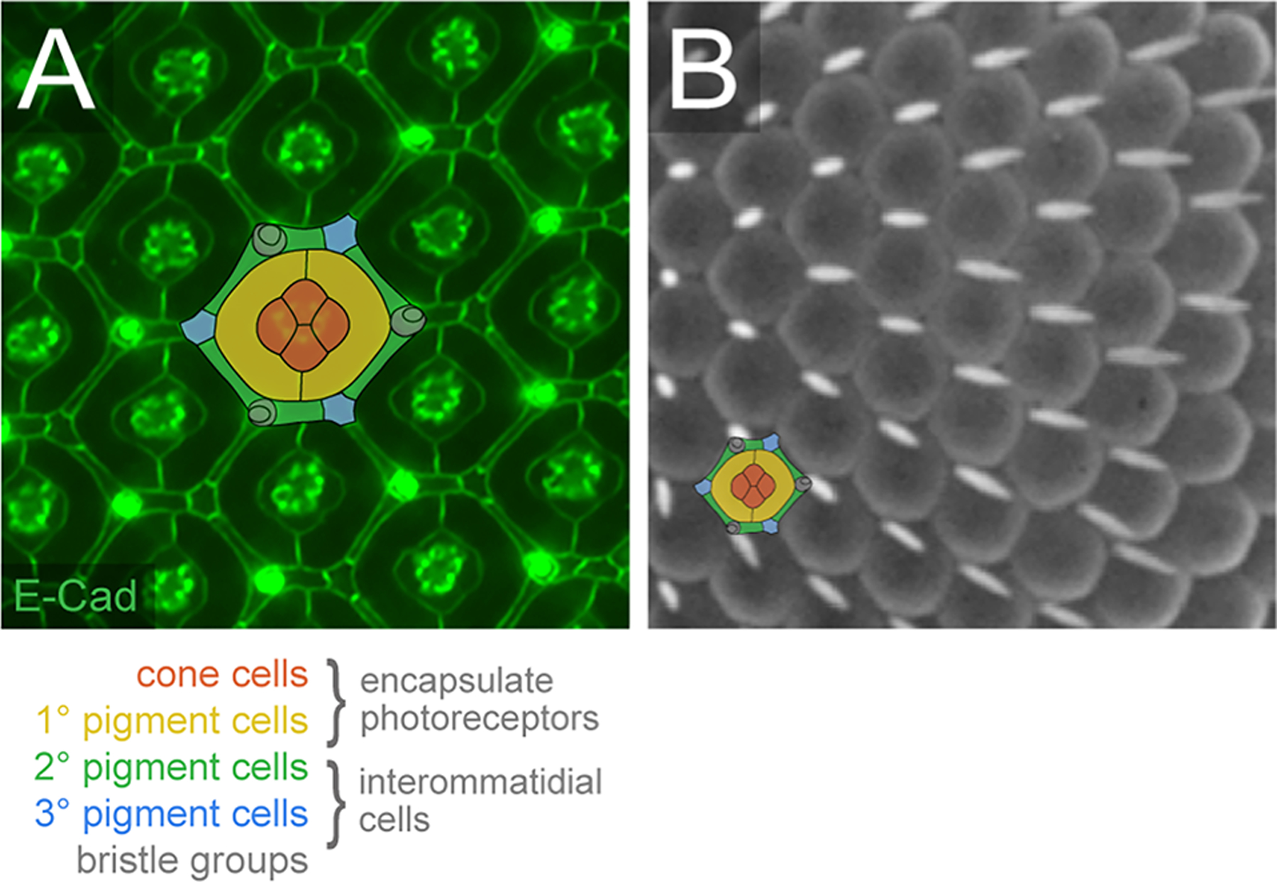
Figure 1: The Drosophila compound eye is highly ordered.

(A) Small region of the pupal eye at 40 h APF. AJs have been detected with antibodies to E-Cadherin. The epithelial support cells are color-coded, as indicated. (B) Small region of a scanning electron micrograph of the adult eye. A single ommatidium is illustrated to emphasize the cells that lie below the rounded lenses, although in the adult eye the IC lattice is more compressed than illustrated. (Images: RIJ)
