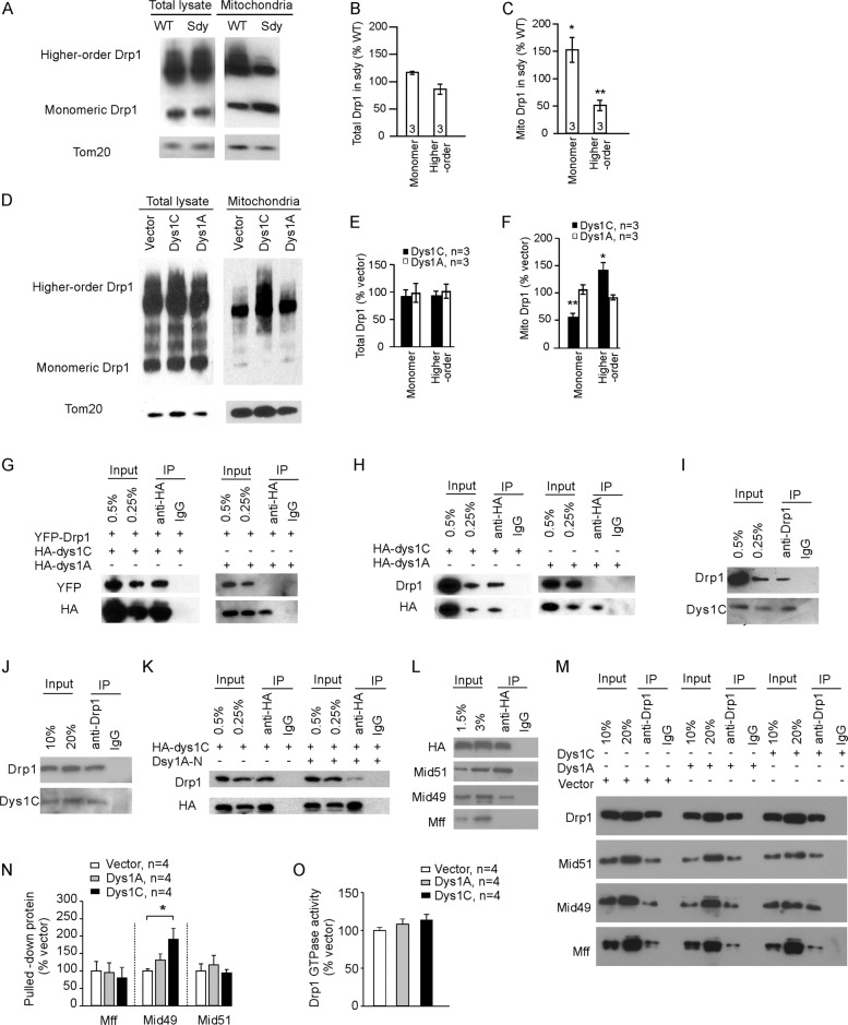
Fig. 4. Dysbindin-1 binds to and promotes Drp1 oligomerization.
Total lysates and the mitochondrial fraction were prepared from the hippocampus of sdy mice and their WT littermates (A–C) or cultured hippocampal neurons transduced with designated lentivirus (D–F), crosslinked and separated by gel electrophoresis. A, D Represent blots. B, C Quantification for A; Student’s t-test was used to compare Drp1 monomer and higher-order structures between sdy and WT mitochondrial fractions; n in the bars indicates the number of mice. E, F Quantification for D; one-way ANOVA was used to compare across groups in E [F(4, 10) = 0.121, p = 0.972] and F [F(4, 10) = 16.371, p < 0.001]; Student’s t-test was used to compare Dys1C vs. vector and Dys1A vs. vector for Drp1 monomer and higher-order structures; n indicates the number of neuronal cultures. G, K, L Immunoprecipitation from HEK-293 cells transfected with designated plasmids using the anti-HA antibody. H Immunoprecipitation from cultured neurons transduced with designated virus using the anti-HA antibody. I Immunoprecipitation from the hippocampus of WT mice using an anti-Drp1 antibody. J Immunoprecipitation from cultured WT neurons using an anti-Drp1 antibody. M Immunoprecipitation from HEK-293 cells transfected with designated plasmids using an anti-Drp1 antibody. N Quantification for M; one-way ANOVA was used to compare across groups for Mid49 [F(2,11) = 4.764, p = 0.039], Holm-sidak was used for post hoc multiple comparisons. O GTPase activity of Drp1. Data are presented as mean ± SEM; *p < 0.05, **p < 0.01.

