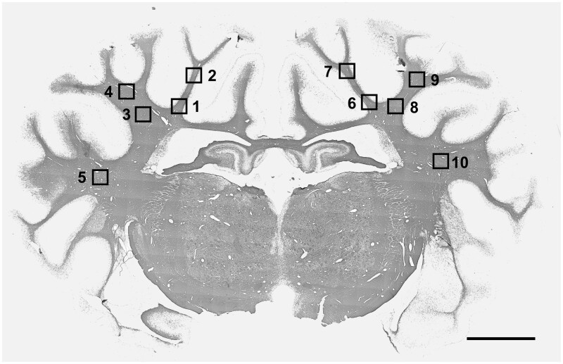
Figure 1.
Photomicrographs showing the fields of view used for analysis of white matter. Photomicrographs showing the fields of view used for immunohistological analysis in the periventricular, parasagittal and lateral white matter. For sections taken at 17 mm anterior to stereotactic zero, one field in the periventricular white matter (squares 1 & 7), and one field in the corona radiata of the first and second parasagittal gyri (2, 4, 8, 10), in both hemispheres, were used for assessment. For Olig2-, 2′,3′-Cyclic-nucleotide 3'-phosphodiesterase (CNPase)- and myelin basic protein (MBP)-labelled brain sections, an additional field was assessed in the upper white matter tract of the first and second parasagittal gyrus (3, 5, 9, 11) and the lateral white matter (6, 12). Section stained with MBP. Magnification ×1. Scale bar is 5 mm.

