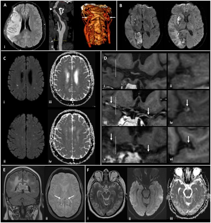
Figure 2.
Magnetic resonance imaging demonstrating the range of neurological complications seen in this study. (A) Territorial infarct, secondary to internal carotid artery (ICA) dissection in a middle-aged previously fit male: Axial fluid-attenuated inversion recovery image (i) showing a right middle cerebral artery (MCA) territory infarct following decompressive craniectomy for malignant MCA syndrome despite treatment with thrombolysis. Reformatted images from a CT angiogram (ii) showing irregularity of the extracranial segment of both internal carotid arteries, consistent with dissection (arrows), with tight stenosis of the true lumen on the right (arrowhead). (B) Multiple territorial infarcts in a female >60 years old with hypertension and dyslipidaemia: Diffusion-weighted images (DWI) demonstrate recent infarcts in the right medial occipital lobe and lentiform nucleus, involving the territories of the right posterior cerebral artery and lenticulo-striate perforators of the right MCA respectively. (C) Acute lacunar infarcts due to small vessel vasculopathy in a male > 60 years old, with a background of hypertension and type 2 diabetes: B1000 images (i, ii) and corresponding apparent diffusion coefficient (ADC) maps (iii, iv) from DWI showing multiple tiny foci of restricted diffusion. (D) Vasculitis in a male >60 years old, with a background of type 2 diabetes, hypertension and hypercholesterolaemia: T1-weighted SPACE vessel wall imaging of both distal ICAs and proximal MCAs, with curved multiplanar coronal reconstructions along the course of both proximal MCAs (first column) and perpendicular to the right MCA (second column, at the position of the dotted line). Pre-treatment pre-contrast (i, ii) and post-contrast images (iii, iv) demonstrate abnormal concentric, long segment vessel wall enhancement (arrows) of both proximal MCAs. Post-contrast images after treatment with prednisolone and tocilizumab (v, vi) demonstrate treatment response with resolution of the previous abnormal mural MCA enhancement (arrows). (E) Acute encephalomyelitis with haemorrhage in a middle-aged male, with a history of chronic obstructive pulmonary disease, who required intensive care and haemofiltration: Coronal FLAIR (i) and axial gradient echo (ii) images showing focal heterogeneous signal abnormality and swelling of the splenium of the corpus callosum, with peripheral low signal indicative of haemosiderin staining (arrows). Confluent high signal is present in periventricular and deep white matter of the parieto-occipital region. (F) Typical imaging appearances of posterior reversible encephalopathy syndrome in a normotensive middle-aged female: Axial T2 image (i) demonstrating hyperintense signal in subcortical white matter of both occipital lobes, with B1000 image (ii) and ADC map (iii) from DWI showing no corresponding restricted diffusion.

