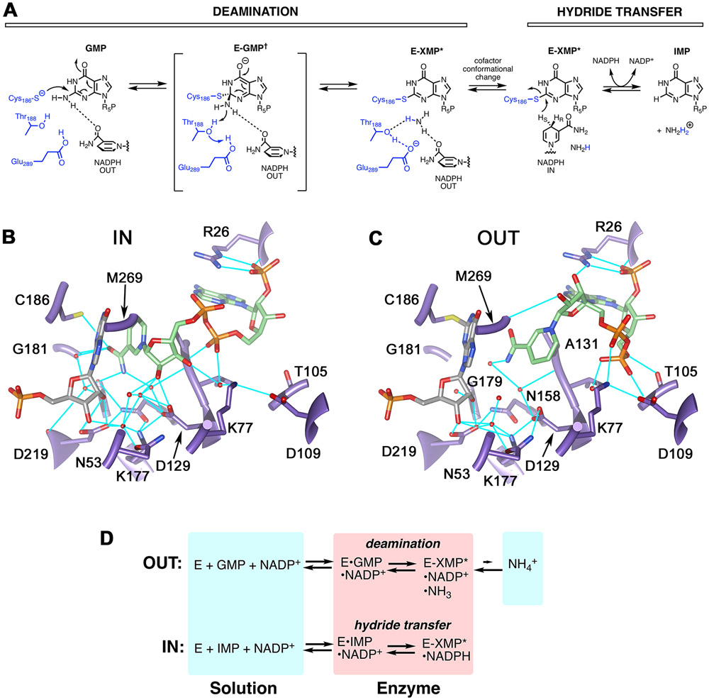
Figure 1. The structure and reactions of GMPR.
(A) The GMPR reaction. (B) The IN conformation as observed in subunit B of 2C6Q 17. (C) The OUT conformation as observed in subunit D of 2C6Q. Protein is shown in purple, IMP is gray, NADPH is green and hydrogen bonds are cyan. The side chain of Met269 has been removed for clarity. E. coli GMPR numbering is used for consistency with experiments. Panels B and C rendered with UCSF Chimera 18. (D) Partial reactions catalyzed by GMPR. Panel adapted from reference 12.

