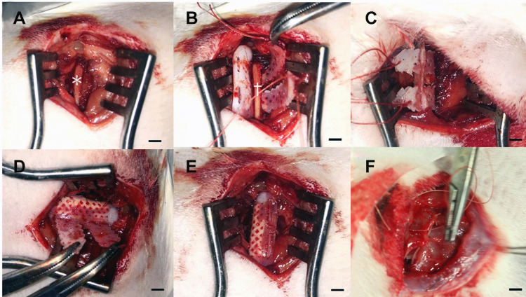
Figure 2.
Rib-fracture model and implantation process. (A) The target rib (*) was reached after careful dissection of the soft tissue layer by layer. (B) An oblique fracture was created (†), and the hybrid fixator was placed underneath the osteotomized rib. (C) and (D) Belts were passed through matching holes and tightened. (E) The over-length belts were cut to smoothen the edges of the fixator. (F) The overlying muscular and skin tissues were approximated layer by layer. Scale bar: 1 cm.

