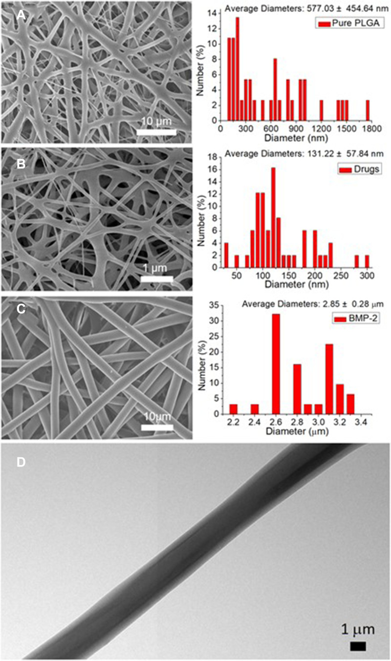
Figure 4.
Scanning electron microscopy images of pure and biomolecule-loaded poly(lactic-co-glycolic acid) (PLGA) nanofibers and their size distributions. (A) Pure PLGA nanofibers, (B) drug-loaded nanofibers, and (C) bone morphogenetic protein-2 (BMP-2)-incorporated sheath-core structured nanofibers. (D) Transmission electron microscopy image of BMP-2-incorporated sheath-core structured nanofibers.

