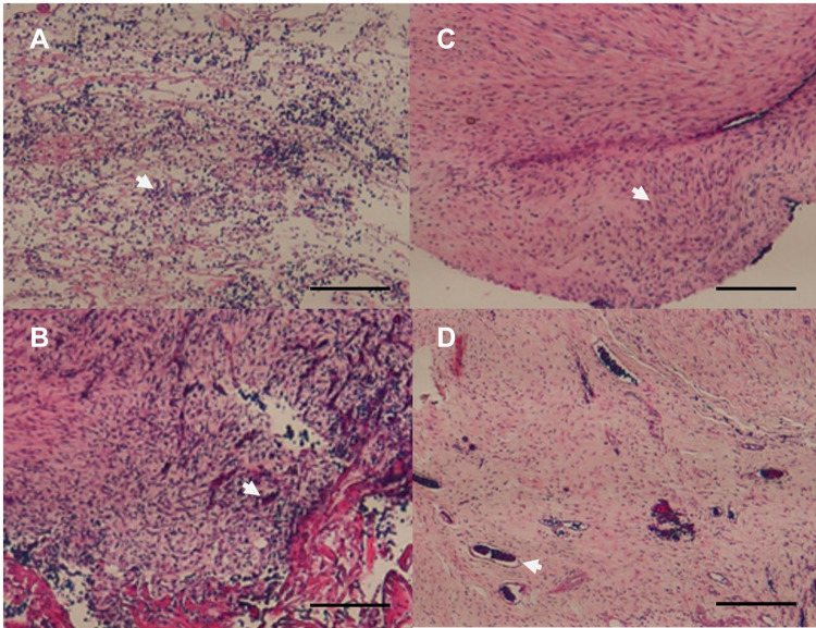
Figure 10.
Histological images (hematoxylin and eosin staining) of the fixator/drug group. Rich mono- and multi-nuclear cells (arrow) were observed at 1 week (A) and 2 weeks (B). Oriented fibroblast-like spindle cells (arrow) could be observed at 3 weeks (C), and several small blood vessels (arrow) were observed at 4 weeks (D) post implantation. Scale bar, 500 μm.

