
Fig. 3.
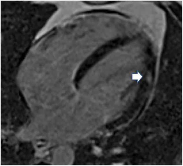
Cardiac magnetic resonance of a four-chamber, long-axis T2-weighted image demonstrating small focal edema (arrow) in the anterolateral wall of the left ventricle.
Official websites use .gov
A
.gov website belongs to an official
government organization in the United States.
Secure .gov websites use HTTPS
A lock (
) or https:// means you've safely
connected to the .gov website. Share sensitive
information only on official, secure websites.
Cardiac magnetic resonance of a four-chamber, long-axis T2-weighted image demonstrating small focal edema (arrow) in the anterolateral wall of the left ventricle.