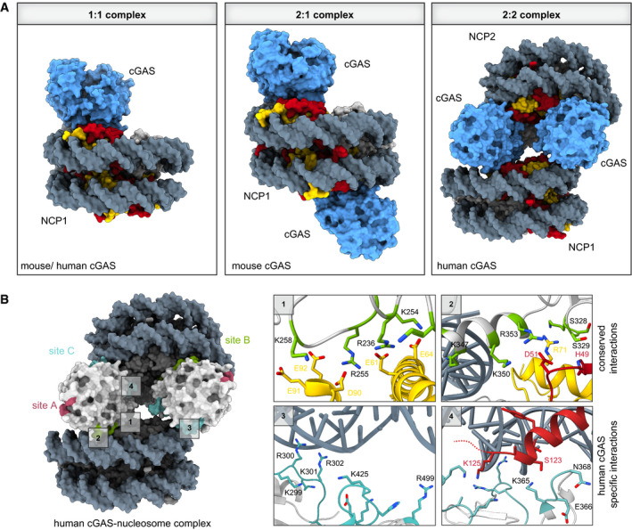
Figure 4. Stoichiometries of mouse and human cGAS–nucleosome complexes and their interaction sites.

(A) Comparison of cGAS–nucleosome complex stoichiometries from different cryo‐electron microscopy structures. While mouse cGAS is found in 1:1 (PDB code 7A08) or 2:1 complexes with NCPs (PDB code 6XJD), human cGAS exhibits a preferred 2:2 stoichiometry (PDB code 7COM). (B) Close‐ups of four distinct cGAS–nucleosome interaction interfaces involving DNA binding sites B and C (PDB code 7COM and 6Y5D). Human cGAS DNA binding site A (pink), site B (green), and site C (cyan) are depicted. Close‐ups 1 and 2 show the conserved residues in site B involved in anchoring cGAS to the acidic patch, and close‐ups 3 and 4 show the human‐specific cGAS site C residues involved in interactions with nucleosomal DNA of NCP2.
