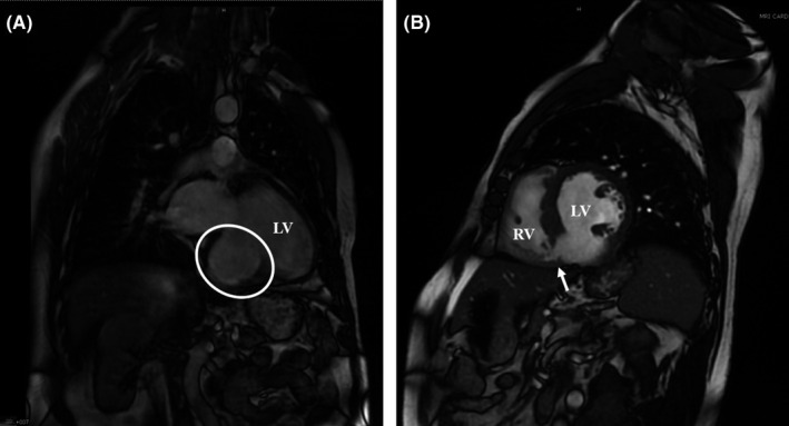
FIGURE 2.

Cardiac magnetic resonance imaging showing left ventricular septal defect and aneurysm. A, Cardiac magnetic resonance imaging (MRI) coronal view showing the left ventricular (LV) inferobasal septal aneurysm (outlined by the white circle). B, Cardiac MRI sagittal view showing the inferobasal septal defect (black arrow) with the interventricular connection between the left ventricle (LV) and the right ventricle (RV)
