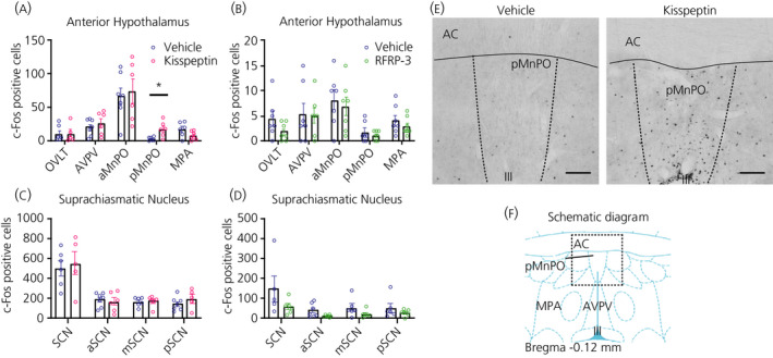
FIGURE 8.

Effect of i.c.v. kisspeptin (Kp) and RFRP‐3 on c‐Fos expression in the male Wistar rat hypothalamus. The number of c‐Fos expressing cells was evaluated in various brain areas (ie, OVLT, AVPV, aMnPO, pMnPO and MPA) of the anterior hypothalamus (A, B) and anterior, medial and posterior parts of the suprachiasmatic nuclei (C, D), 60 min after the i.c.v. injection of Kp (3 nmol), RFRP‐3 (250 pmol) or vehicle (NaCl 0.9%). (E) Showing representative images of c‐Fos staining in the median preoptic nucleus (pMnPO) of animals injected with vehicle (left) or Kp (right). (F) Showing a schematic diagram obtained from the rat brain atlas (Paxinos and Watson) of the preoptic nuclei at a level equivalent to bregma −0.12 mm.21 Data represent the mean ± SEM of n = 5 ‐ 7 animals, with scattered dots representing individual values per animal. *P < 0.05 statistical difference between Kp and vehicle after Student's t‐test. III, third ventricle; AC, anterior comissura; aMnPO, anterior part of the median preoptic nucleus; AVPV, anteroventral periventricular nucleus; MPA, medial preoptic area; OVLT, organum vasculosum of the laminae terminalis; pMnPO, posterior part of the median preoptic nucleus; SCN, suprachiasmatic nucleus. Scale bar =200 µm
