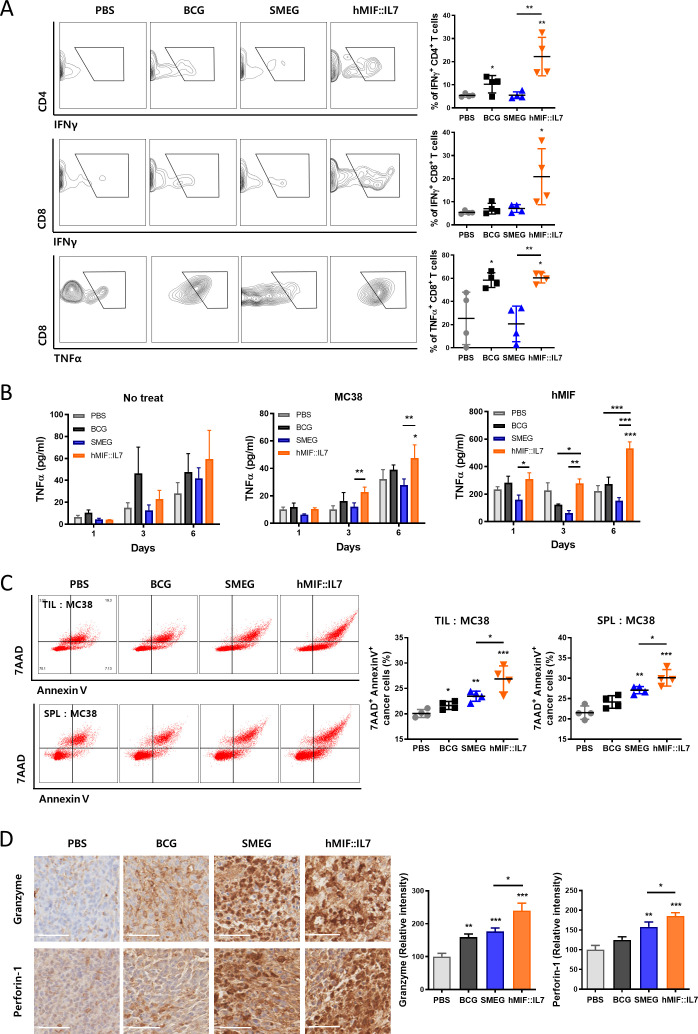
Figure 3.
rSmeg-hMIF-hIL-7 induced antitumor immune responses through the recruitment of functional T cells in the tumor environment. (A) The population of IFNγ-releasing CD8+ T cells and CD4+ T cells and TNFα-releasing CD4+ T cells in tumors were analyzed by flow cytometry. (B) Splenocytes were incubated with MC38 lysates or h-MIF for 6 days. Inflammatory cytokine secretion of splenocytes in response to the antigen stimulation was detected by ELISA. (C) Four days after the last mycobacteria injection, lymphocytes from the tumor tissue and spleen were isolated and cocultured with carboxyfluorescein succinimidyl ester (CFSE)-labeled MC38 (E:T=5:1) cancer cells, and cytotoxicity of the lymphocytes were detected by apoptosis assay. (D) IHC image for cytolytic responses by secretion of the pore-forming molecule perforin-1 and proapoptotic protease granzyme B in tumor tissue (scale bar=50 µm). Significance differences (*p<0.05, **p<0.01, and ***p<0.001) among the different groups are shown in the related figures, and the data are presented as the mean±SEM of mice (n=4).

