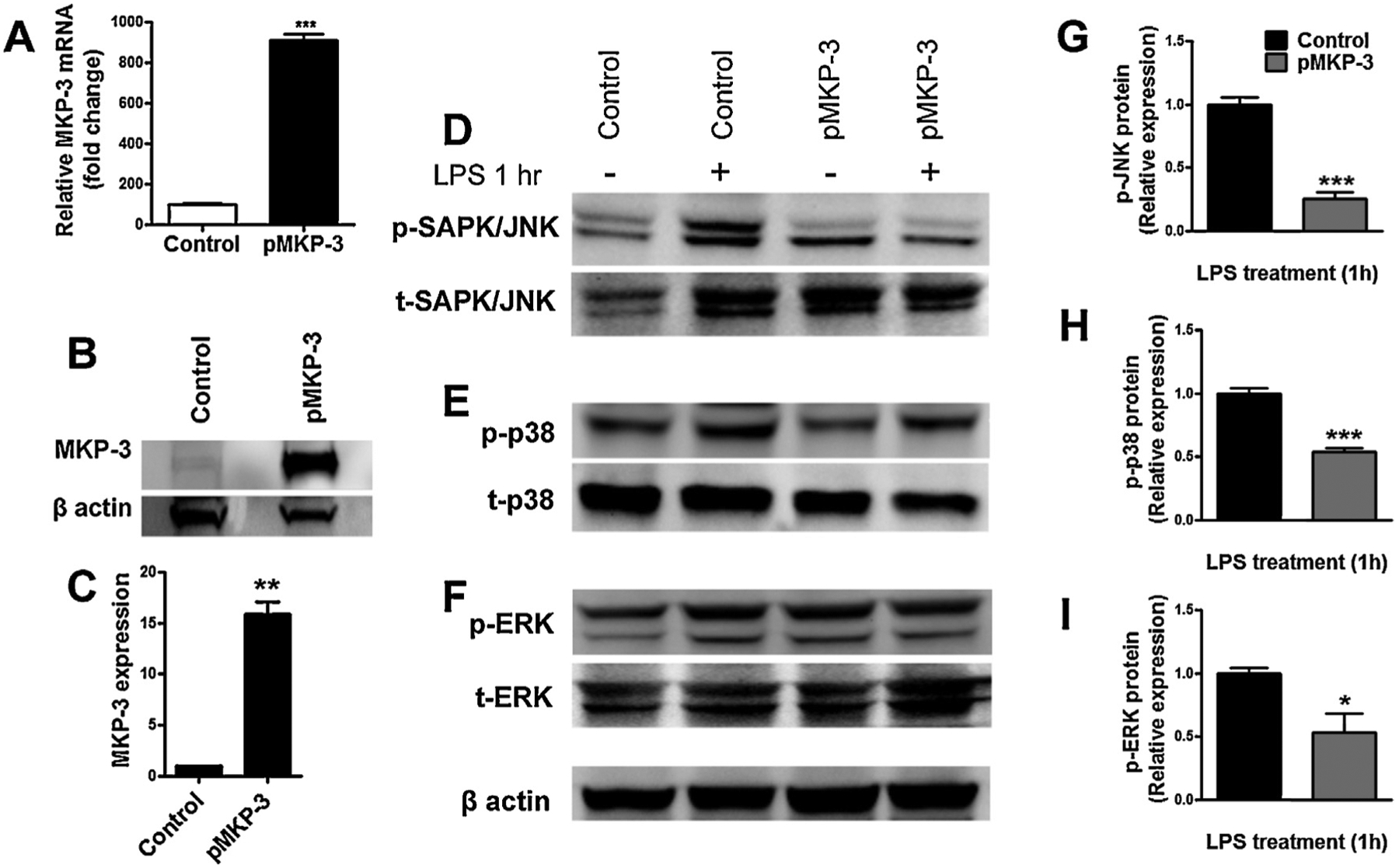
Fig. 1.

In vitro characterization of MKP-3 and MAPKs in primary astrocytes cells transiently expressing MKP-3. MKP-3 mRNA levels determined by qRT-PCR (A), representative Western blot (B) and quantification of MKP-3 protein (C) in normal primary astrocytes (control) and primary astrocytes transiently overexpressing MKP-3 (pMKP-3). Representative Western blots of p-JNK (D), p-p38 (E), and p-ERK (F), and quantification of p-JNK (G), p-p38 (H), and p-ERK (I) protein in normal primary astrocytes (control) and primary astrocytes transiently overexpressing MKP-3 (pMKP-3), following 1 h incubation in medium (control alone) or lipopolysaccharide (LPS) stimulation (1 μg/ml). Results are expressed as mean ± s.e.m. of three experiments in triplicate. * P < 0.05, ** P < 0.01, *** P < 0.001 vs. control primary astrocytes.
