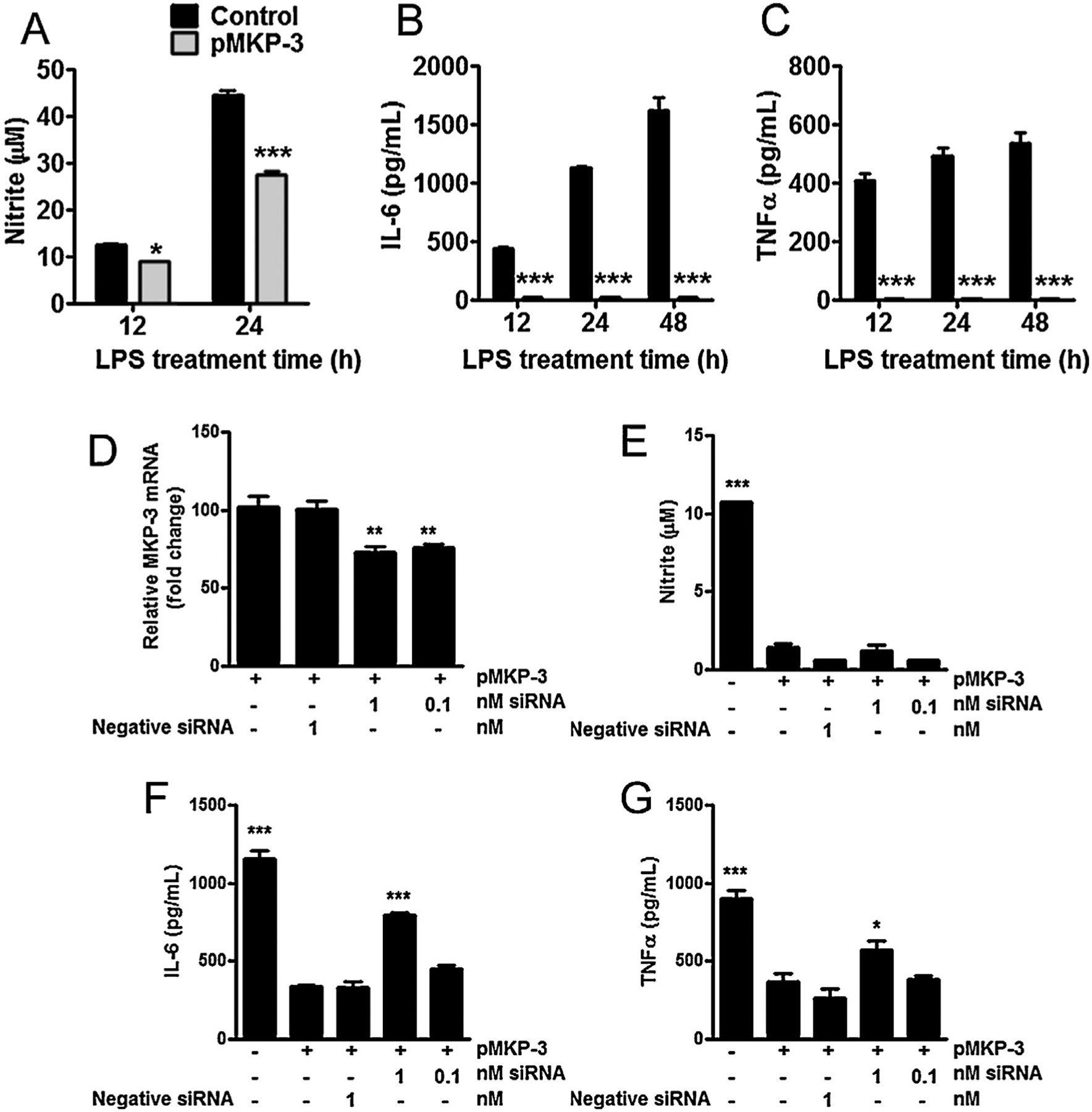
Fig. 2.

Overexpression of MKP-3 reduces inflammatory effectors in primary astrocytes cells and reversal effects of MKP-3 mRNA knockdown in BV-2 microglia cells. (A) Nitrite oxide (measured as nitrite), (B) IL-6 and (C) TNFα concentration in cell culture supernatant in control (normal) and transiently expressing MKP-3 primary astrocytes cells challenged with LPS (1 μg/ml) for 12, 24 or 48 h. MKP-3 mRNA levels (D) in BV-2 cells stably expressing MKP-3 after 24 h transfection with MKP-3 siRNA compared to negative control siRNA and vehicle treated cells. Nitrite oxide (measured as nitrite) (E), IL-6 (F), and TNFα (G) cytokines release in control (normal) and stably expressing MKP-3 BV-2 cells transfected 24 h with a negative siRNA or a siRNA against MKP-3 and challenged with LPS (1 μg/ml) for 24 h. Results are expressed as mean ± s.e.m. of three experiments in duplicate. * P < 0.05, ** P < 0.01, *** P < 0.001 vs. control primary astrocytes or BV-2 cells stably expressing MKP-3.
