
FIGURE 1.
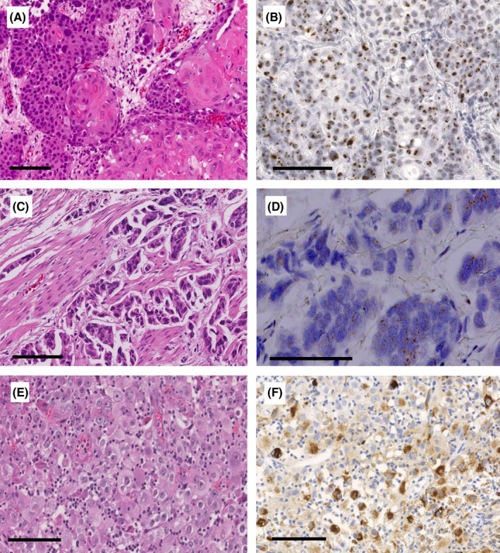
Representative light microscopy images of HPV‐associated urothelial carcinoma (UC). An invasive UC with squamous differentiation (HE stain, A), showing nuclear positivity for high‐risk HPV E6/E7 mRNA by ACD RNAscope® Probe HPV HR18. (RISH stain, B) Muscle‐invasive micropapillary UC (HE stain, C), showing positive brownish punctate staining in the cell nuclei and cytoplasm for high‐risk HPV E6/E7 mRNA by RNAscope (RISH stain, D). An invasive high‐grade UC (HE stain, E), showing strong cytoplasmic immunohistochemical reaction for a major HPV capsid antigen (IHC stain, F). Scale bars represent 100 μ m