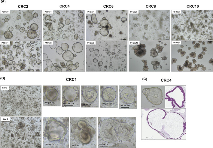
FIGURE 2.

PDOTS morphology in bright‐field microscopy. Multicellular OTS derived from colorectal cancer patients were observed in the phase‐contrast imaging. (A) PDOTS morphology remained the same cystic and/or luminal structures in both Passage 0 and Passage 4. (B) The size of the CRC1 PDOTS was approximately 100 μm following 2 days of culture; they increased in size by twofold within 5 days (C) H & E staining of OTS from CRC4 patient tumor tissue. Scale bar, 100 μm
