FIGURE 4.

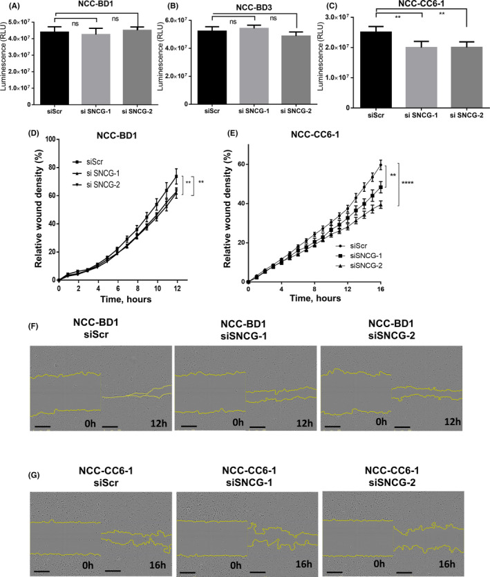
Cell proliferation and wound healing assays. (A–C) Cell proliferation assays were performed with gamma‐synuclein (SNCG)‐overexpressing cell lines NCC‐BD1, NCC‐BD3, and NCC‐CC6‐1. Cells were seeded onto 96‐well plates. After 24 h, siRNA was added, and, after a further 72 h, cell viability was measured. SNCG knockdown by siRNA against human SNCG (siSNCG‐1 and siSNCG‐2) had no effect on the proliferation of NCC‐BD1 or NCC‐BD3 cells compared to si‐negative control‐treated cells (siScr), but SNCG knockdown did suppress the proliferation of NCC‐CC6‐1 cells. Each assay was replicated in three wells. Shown are the means ± SDs of representative data from three independent assays. p values were calculated using Student’s t‐test. **p < 0.01. (D–G) Cell migration was assessed by wound healing assays using an IncuCyte Zoom Kinetic Live Cell Imaging system. Cells treated with siRNA were plated in six replicates into 96‐well plates coated with collagen I. Four hours after seeding, scratches were made. SNCG knockdown downregulated the migration of NCC‐BD1 (D) and NCC‐CC6‐1 (E) cells significantly compared to si‐negative control‐treated cells. Data plots show the means ± SEs. p values were calculated using Student’s t‐test at 12 h after making the scratch for NCC‐BD1 cells and at 16 h after making the scratch for NCC‐CC6‐1 cells. **p < 0.01, ****p < 0.0001. Representative figures showing wound healing of NCC‐BD1 (F) and NCC‐CC6‐1 (G) cells. Scale bars indicate 300 µm. The graphs and pictures are representative graphs of three independent experiments
