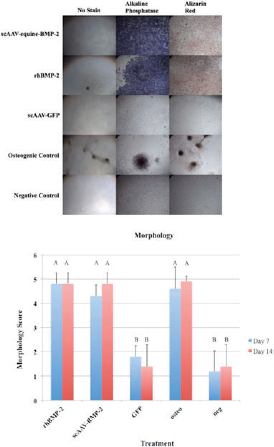
Figure 2.

(a) MSCs in culture treated with scAAV-equine-BMP-2, rhBMP-2, and osteogenic media exemplify osteogenic morphologic changes at day 7 following transduction. (b) Morphology Scores: Error bars denote standard deviation from the mean. Letter changes denote significant differences between groups. ScAAV-equine-BMP-2 transduced cells, rhBMP-2 treated cells, and osteogenic control cells appeared significantly more osteogenic than GFP transduced cells and negative controls (p < 0.0001). The morphological changes were evident by day 7.
