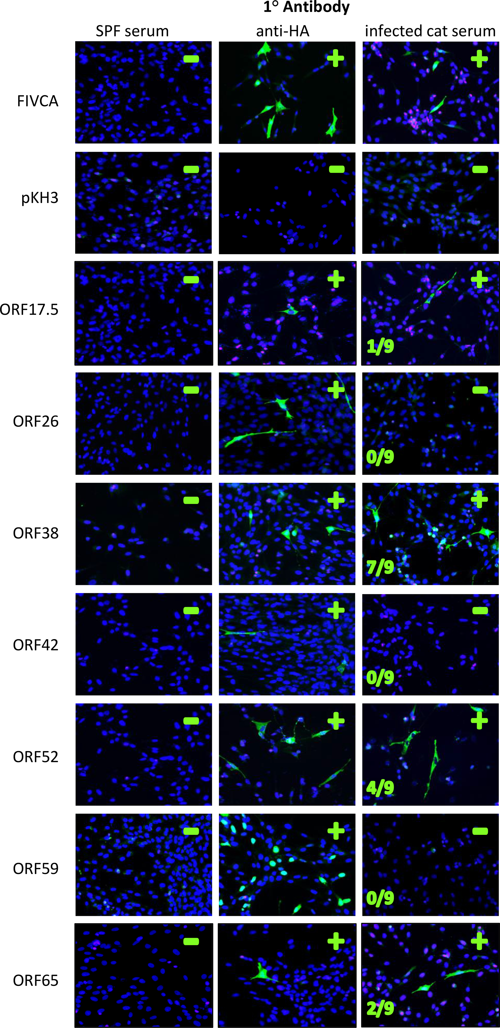
Figure 1. IFA detects FcaGHV1 antibodies in infected cats.

The left protein antigen that was transfected into each set of cells. SPF-naive cat serum was used as a Fluorescence in the second column (anti-HA) indicates cells expressing the protein of interest with an HA tag. Fluorescence in the third column indicates cells that bound cat serum antibody. Nuclei are stained with DAPI and appear blue. Serum from nine FcaGHV1-qPCR positive cats was used to screen each transfection reaction to determine if antibodies were present in cat sera for each antigen tested, number of cats positive is indicated. Representative results with serum from one FcaGHV1 qPCR-positive cat are shown here, see Table 2 for tabulation of these results. The FIV capsid (FIVCA) was used as a positive control for detection of viral antigen when exposed to sera from FIV+ cats. A vector-only negative control, pKH3, was also run with each transfection. FIVCA= FIV capsid protein.
