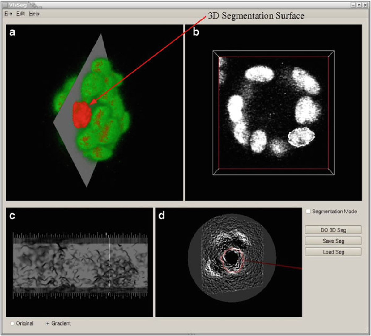
Fig. 10.

3D Segmentation. (a VisSeg 3D volume rendering window showing the segmentation surface after completion of 3D segmentation of a nuclei. (b) 2D slice showing the 2D segmentation in a specific slice. (c) 3D segmentation evaluation window showing the segmentation quality. (d) 3D segmentation correction window where additional correction points can be added
