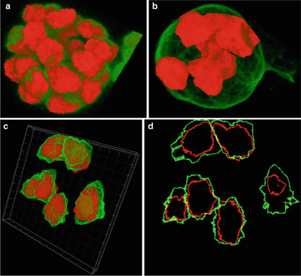
Fig. 11.

Segmentation correction. (a) Segmentation of all nuclei in the MCF-10A sample using volume staining channel. (b) Partial segmentation of whole cells in the MCF-10A sample using surface staining channel. (c) Partial segmentation of the somite stage mouse embryo sample with surface staining visualized with Imaris (http://www.bitplane.com/go/products/imaris). (d) Slice 13 of the mouse embryo sample showing the nuclear (red) and surface (green) segmentation
